A Mysterious Death in a 21 yo Healthy White Female, and the Larson Maneuver

My wife is at work at a hand surgery pre-op care clinic. It is her birthday. In walks a middle-aged male who is about to have tendon reconstruction after he sustained a crush trauma, and is excited about the possibility of returning to work. He is slightly abrasive and somber, despite the prospect of receiving the surgery.

My wife asks him if he is concerned about the procedure, asks if he is feeling well, asks if there is anything she can do to help. With a quiet manner he states all is well. To make discussion my wife states today is her birthday and she is excited to celebrate when she gets home. He smiles and states today is also his daughter’s birthday. She was born the same year as my wife. He states today is always a rough day for him because she passed away in an emergency room 7 years ago at 21 years of age.

We can never fully understand where those we treat are coming from, their life experiences, or what their home situations, thoughts, fears, dreams, and worries entail; and this was a reminder for me. He stated that his daughter was healthy,woke up without issue on that day, but later developed difficulty breathing and wheezing. She was diagnosed with an acute asthma attack. She had one episode in the distant past but was not on any routine medications. She was given albuterol and experienced a negative reaction to the albuterol and completely stopped breathing. She was brain dead by the time she was intubated. They withdrew care in the emergency department.

After my wife shared this with me, I searched to find any case reports of paradoxical reactions to albuterol. Below are three related cases, however bronchospasm becoming worse with beta-blockers is exceptionally rare.

Case reports of paradoxical bronchospasm to inhaled beta agonists:

What I think more probable, and possibly related to the above case reports, is acute laryngospasm. The albuterol she received may have further irritated her vocal cords potentially worsening, rather than relieving her vocal cord dysfunction. Both Resus.me and LITFL (Life In The Fast Lane) have very useful articles describing management (see below for links). Here is a brief synopsis:

Laryngospasm

What is it?: a potentially life-threatening closure of the vocal chords (can occur spontaneously). Often misdiagnosed as asthma—especially exercise-induced asthma (more common in white females).

How to diagnose (and differentiate from asthma):

  • Stridorous sounds are usually loudest over the anterior neck, beware wheezing sounds transmit throughout the lungs
  • Typically, albuterol has minimal to no beneficial effect.
  • Subjectively more difficulty on inspiration than expiration

   Clues in history: recent exercise, GERD, ENT procedures, or extubation

   Common causes & some that are not-so common:

  • Post extubation
  • Exercise
  • GERD
  • Medications (e.g., (1) ketamine sedation, incidence 1-2 %; (2) versed (very rarely), which can be reversed with flumazenil)
  • Near drowning/ aspiration
  • Inhalants (smoke, ammonia, dust, cleaning chemicals)
  • Related to anxiety
  • Strychnine (plant based poison, sometimes used as a pesticide for birds and rodents, also the poison reportedly used to kill Alexander the Great in 323 BC)

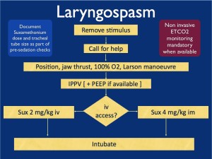
Treatment of laryngospasm:

Initially:

  1. Jaw thrust with Larson Maneuver
  2. CPAP/ NIPPV
  3. Heliox might be helpful if available, (also topical lidocaine can be applied to larynx if available)

If conservative measures fail:

  1. Low dose propofol (0.1 mg/kg) ~ give 10 mg
  2. Low dose succinylcholine (AKA: suxamethonium) 0.1-0.5 mg/kg IV
  3. All else fails: intubation with succinylcholine 1.5 mg/kg IV
    • If no IV access, then succinylcholine IM (3-4 mg/kg). Experts advocate IM injection into the tongue.
    • Perform chest thrust maneuver immediately preceding intubation to open the vocal cords and allow passage of the ET tube.
    • Monitor for negative pressure pulmonary edema—(from patient pulling hard against closed glottis in the setting of acute asphyxia).

Flow chart from Resus.me

Larygospasm_flow_high_res

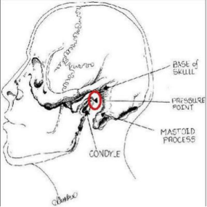
What is the Larson Maneuver? (Published 1998 in Anesthesiology)

It is a manipulation jaw thrust technique targeted at the ‘Larson’s point‘, AKA: laryngospasm notch.

  • Place middle finger of each hand in the laryngospasm notch, located behind the lobule of each ear, between ascending ramus of the mandible and the mastoid process.
  • Press very firmly inward toward the base of the skull with both fingers
  • At the same time lift the mandible at a right angle to the plane of the body (perform jaw thrust).

Reportedly will convert laryngospasm within one or two breath cycles to laryngeal stridor, and in after a couple more breath cycles, to unobstructed respirations. As proposed by Larson, it is likely that the painful stimulus relaxes the vocal cords by way of either the parasympathetic or sympathetic nervous systems through the glossopharyngeal nerve.

Diagram from LITFL

Larson_man

References:

  1. Resus.Me: http://resus.me/laryngospasm-after-ketamine/
  2. LITFL (Life In The Fast Lane): http://lifeinthefastlane.com/ccc/laryngospasm/
  3. UpToDate: https://www.uptodate.com/contents/paradoxical-vocal-fold-motion?source=machineLearning&search=Laryngospasm&selectedTitle=1~150&sectionRank=1&anchor=H3#H3
  4. Larson, Philip, MD. Laryngospasm-The Best Treatment. Anesthesiology. 1998. http://anesthesiology.pubs.asahq.org/article.aspx?articleid=1947036
  5. Paradoxical bronchospasm: a potentially life threatening adverse effect of albuterol. South Med J. 2006 Mar;99(3):288-9. http://www.ncbi.nlm.nih.gov/pubmed/16553105
  6. Paradoxical response to levalbuterol. J Am Osteopath Assoc. 2008 Apr;108(4):211-3. http://www.ncbi.nlm.nih.gov/pubmed/18443029
  7. Paradoxical reaction to salbutamol in an asthma patient. Pneumologia. 2012. Jul-Sep;61(3):171-4. http://www.ncbi.nlm.nih.gov/pubmed/23173379

Hypotension

Late 70s year old female with chief complaint of dizziness and fatigue. Patient has a medical history of HTN and recent cataract surgery.  In triage patient was hypotensive with BP of 70\35 and Bradycardic with a heart rate ranging from 55-60, O2 sats 100% on room air and afebrile.

I found the patient to be lethargic. Otherwise her exam was unremarkable with no focal neurologic deficits, cardiac and pulmonary exam unremarkable, and no abdominal pain. Her husband was in the room and states that they were on their way to their grandson’s high school graduation and she began complaining of feeling dizzy and she started to become lethargic. She had not been sick recently and before this morning she was completely at her baseline. To note yesterday her blood pressure was 180/95 when she checked at home so she typically runs high. No new changes to her medications which consisted of metoprolol Succinate 100mg QD and a Baby Aspirin.

As I talked to the husband he went on to explain that she had recently had cataract surgery on her left eye and that this morning she had a follow up appointment with her ophthalmologist. While at the ophthalmologist appointment the doctor said that the pressure in her eye was high and she received some drops in her eye to bring the pressure down but the husband could not remember the name of the drops.

So I started by getting CBC, CMP, TSH, Urine, Chest xray, EKG, Troponin and a head CT to complete my little old lady AMS workup. Obviously the differential diagnosis for AMS in the elderly is vast so I was considering a lot of different possibilities.  While I waited for her results to come back I called her Ophthalmologist that she had seen that morning to see what drops she had received. Ends up she got Alphagan which is an alpha agonist, Trusopt which is a carbonic anhydrase inhibitor, and 3 drops of Timolol. I discussed with him the possibility of the Timolol on top of her morning metoprolol 100mg as potentially causing her hypotension and bradycardia. He stated he had never seen that happen in his 16 yrs of practice but it is theoretically possible. A quick Uptodate search confirmed that hypotension can occur in as many as 10% of patients using Timolol eye drops which to me was a surprisingly high number.

So I’ll go ahead and cut to the good stuff. All of her labs and imaging returned unremarkable and her EKG just showed sinus bradycardia with a rate of 57.  Ultimately she got 2L of Normal Saline and we watched her for about 5 hrs. Throughout her stay her blood pressure steadily increased and at the time of her discharge she was 135/86 with a heart rate of 74 and she was back to her baseline and much more awake.

I thought this was an interesting case as it seems relatively rare for topical eye drops to result in systemic side effects however in the right patient population it can result in severe side effects. A quick literature search brought up multiple case reports of patient’s having symptomatic bradycardia and even syncope resulting from Timolol use.

So definitely something to keep in mind if you have a elderly or frail patient with acute angle glaucoma who is already on beta blocker therapy. Maybe trying other drops first instead of Timolol or at least be sure to make the patient aware of the possibility of side effects including hypotension, bradycardia, fatigue, and even syncope so they know what to watch out for.

Sick PEs

We had a very ill patient recently. She was found down upstairs visiting her family member. She was calmly altered, not agitated but was in mild distress. Consciousness fluctuated. Tachycardic and hypertensive initially, then had more labile BP and some hypotension.

We had to intubate her due to poor MS and clinical condition. She coded in CT, we placed a central line on the CT table between her noncontrast head CT and her CT chest. We pushed an amp of Epi and ran out of the room for the CT chest. We were worried about dissection and PE in equal amounts. We could not get good cardiac windows on bedside Echo in room 9 prior to the CT.

She continued to intermittently lose her pulse and drop her BP. We confirmed bilat PEs on the CT when we saw NO contrast left her right ventricle. The CT tech noticed first and became worried the patient had no cardiac output (ie pulseless).

We rushed the patient back to room 9 and gave a tPA bolus (50mg) followed by infusion of 40mg. She was on pressors and heparin and improving. Dr Smith accepted her to Jewish for possible EKOS or even ECMO if needed. On arrival to Jewish a few hours later she coded and died.

I was surprised when I found out she had died. Her O2 sat was improving, HR was decreasing, blood pressure was stable (though dependent on pressors). She received a large amount of crystalloid IV which according to some data might not have been optimal management. She also had the following ECG:

FullSizeRender

I think she was infarcting her myocardium. She likely had pulmonary infarction considering her poor oxygenation. She had coded a few times. She had a lot of strikes against her. Her BEST SHOT was going to a place with catheter assisted treatment for PE and ECMO if needed.

I am posting the case to let everyone know:

  1. How to manage sick PE patients (see post below)
  2. To use tPA in massive and in many cases of submassive PE
  3. TRANSFER sick PE patients to Jewish for EKOS/ECMO
  4. The decision to diagnose PE with RV strain on BEDSIDE Echo with no formal Radiologic testing will depend on your attending

This post from EmCrit / PulmCrit is a beautiful summary with potential dogmalysis related to PE management (see take home points below but do read the post).

In addition, here is a nice review article on catheter-based reperfusion treatment for PE with nice references for further reading.

Take home points from the Emcrit post:

  • The only evidence-based intervention that seems to improve mortality in massive PE is thrombolysis.   The primary goal of therapy should be administration of thrombolysis as soon as possible to patients without contraindication.
  • Consider early stabilization of blood pressure using a norepinephrine infusion, administered peripherally if necessary.
  • Volume administration may facilitate dilation of the right ventricle and hemodynamic deterioration.
  • Intubation is very hazardous and should be avoided if possible.   Patients die from cardiovascular collapse, and intubation may worsen this.
  • For a coding PE patient consider 50mg alteplase bolus as well as an infusion of epinephrine.  Patients can do well despite requiring CPR and high dose vasopressor infusions.

Phrenic Nerve Paralysis after intrascalene nerve block

This was covered in the opening of the February EMRAP however not something that I was at all familiar with.  Here are the basics and something that is probably rare but worth knowing about:

 In the episode they presented a case of  70 year old female that was brought in with a complaint of shortness of breath. RR = 28 and SA02 – 88%. The patient presented three hours after a right shoulder arthroscopy.  

ECG : normal

CXR: elevation of the right hemidiaphragm

 Dx:  paralysis of right hemidiaphram after intrascalene nerve block

There are two major complications associated with intrascalene nerve blocks: 1. pneumothorax

2. unilateral phrenic nerve paralysis.

The patient likely had a transient phrenic nerve dysfunction causing unilateral diaphragmatic paralysis.  Younger patients can compensate, older patients with co-morbid conditions may not be able to tolerate this as only one lung is effectively ventilating.

The patient in this case was managed with supplemental 02 until the buvipicane wore off.  Some patients with underlying lung disease and this complication may require BiPAP/CPAP or intubation.  

Things that I took away:

  •  be aware of this procedure and this complication
  • this may be done for patients with same days surgeries to the upper extremity and this is important history to have from Pt or family.
  • It may be missed prior to d/c.  This was an example of one that was missed by Anesthesia prior to d/c of the patient. 
  • May look clinically like PE (tachypnea, tachycardia, post-op patient), however,  history will help make the dx as well as CXR.


A little more about the block:

Interscalene nerve block is typically performed to provide analgesia for upper extremity surgeries and may or may not be combined with mild general anesthesia.

 Example of Surgeries this may be used for:

-Shoulder surgery, such as rotator cuff repair, acromioplasty, hemiarthroplasty, and total shoulder replacement

– Humerus fracture

Dr Huecker Stayin’ Healthy

As I was catching up on my weekly EM/pharmacology blogs, I stumbled across an article featuring our very own Dr Martin Huecker on the Academic Life in Emergency Medicine (ALiEM) website.  It is a great article about staying healthy and balancing life in EM. I’ve attached the link for your viewing pleasure.

https://www.aliem.com/2016/martin-huecker-healthy-in-em/

Spice/Heroin Reactions

So I’ve had to encounter my two sickest patients in the holding area within the past 2 weeks or so. One was a reminder from intern year while looking through spice charts, while the other was an actual patient that I had 3 days ago.

We always tend to take the “Tank” patients lightly and overlook them sometimes. I just want to use this as a warning (especially to the interns) that sick patients can also use heroin/ETOH/Spice, so pick up on the small interactions that don’t go right. I’ll try to present these starting with how their chief complaint lead to the final diagnosis.

Patient 1 (intern year)

  • middle aged male
  • CC: Spice OD, Nausea/Vomiting
  • Final Diagnosis: Subarachnoid Hemorrhage
  • Time to Diagnosis: 7 hours

So this guy presented as a spice reaction. This was before spice became as widely spread as it is now, and no one knew what to expect symptomatically (not that there is ANYTHING that is characteristic to spice anyways).

General story from talking with this guy is that he used spice for the first time that night. No significant past medical history. He was on a first date where everything had been going well. He had borrowed some spice from his friend where he used back at his place after dinner. Soon after he started having nausea and projectile vomiting and was acting ‘goofy.’ At that his date called EMS and the date ended.

Exam:

  • Gen: Fully A&O, slightly odd in that he seems incredibly happy to be here
  • CV: RRR
  • Pulm: CTAB
  • ABD: NT/ND
  • Neuro: CN II-XII intact, motor intact, sensation intact, ambulates without difficulty to bathroom

This man was like most of our intoxicated patients–a sober re-evaluation. At approximately 2 hours he was still vomiting in the ED, so the medical workup was initiated. Due to his odd behavior with vomiting, we got a CMP/CBC/Tox & CT Head. The night continued busy and I almost forgot about him as I waited for results. Ultimately he never got his CT Head due to being uncooperative but I wasn’t told until hours later. He ended up getting Geodon/Ativan in the ED but instead of calming him down he became more agitated and was no longer oriented. Ultimately getting rolled into room9 to be intubated prior to CT and the final diagnosis was made.

Certain forms of spice that lead to agitation also lead to spikes in blood pressure, and there are a few case reports of significant hypertension occurring after spice use. This guy had the unfortunate case of rupturing an aneurysm after using spice likely from a BP spike. I’m honestly not sure if the outcome would have been any different had I reached the diagnosis sooner — he got repetitive Head CTs and ultimately an EVD on hospital day 3. I didn’t really take him seriously even after I ordered a lab workup. This really changed my perspective on patients being held for intoxication. He spent 1.5 months in the hospital (1 month intubated) before being discharged to rehab.

Patient 2 

  • Middle aged white female
  • CC: Heroin OD got Narcan
  • Final Diagnosis: Cardiogenic Shock
  • Time to Diagnosis: 3.5 hours

This case I handled a bit better (I’d hope after 2 years). Story I could get is that this man had a syncopal episode. Received Narcan PTA by EMS and woke up. In the ED the patient adamantly denies heroin use–states he simply passed out. Luckily I got to him before EMS left, and EMS confirmed reports of bystanders stating opiate use.

Exam:

  • Vitals: HR 120, RR 16, O2 96%, BP 80/45, T 98.0
  • Gen: Fully A&O, drowsy
  • CV: tachycardia
  • Pulm: CTAB
  • ABD: NT/ND
  • Neuro: CN II-XII intact, motor intact, sensation intact, ambulates without difficulty to bathroom

My initial thought was that he may need some more narcan or that he received a longer acting opiate. The tachycardia was a wild card and didn’t make much sense with the picture. He remained afebrile and temp recheck, so I wasn’t thinking sepsis much at that time. At this point due to the tachycardia not making sense I ordered labs (and a tox for co-ingestants) and thought his BP/HR would improve with fluids.

I reassessed him after bolus #1 and #2 and neither HR or BP improved. Labs returned with an elevated WBC at 19.6. Opiates positive on top but otherwise were unremarkable. EKG sinus tachycardia. CXR and urine unremarkable. At this point even though I had no fever or source I felt compelled to initiate a septic workup and Lactate returned at 7.9.

I was starting to get lost as why this guy was so unresponsive to fluids and O’Brien and I threw the USN to bedside at this point. Turns out he was in acute systolic failure with an ejection fraction of 11%. No history of CHF and also no signs of volume overload on exam except very mild pulm edema. Troponin peaked at 0.5.

He was admitted by cardiology while they trended his status. He went to the cath lab on hospital day 3 with clean coronary arteries. Ejection fraction improved to 60% by time of discharge. Talking with the team today they are still uncertain of the cause.

These are two cases of sick patients being in the holding area. Hopefully, it serves to remind everyone that any patient can be sick.

Interesting Ultrasound

A late 20s F G4P3003 at approximately 6 weeks gestation by LMP presents with a chief complaint of vaginal bleeding. A few hours PTA, patient states she felt a “gush of blood” with some mild abdominal cramping. VSS. On exam, noted to have a moderate amount of vaginal bleeding per the os. On our bedside ultrasound we note what appears to be a viable IUP with cardiac activity.  However, the uterus appears septate, with half containing the IUP and the other half more hyperechoic/solid in nature. We were concerned for a possible subchorionic hemorrhage and consulted OB/GYN. Our bedside US image is below:

BS US

OB came down with their Cadillac ultrasound and confirmed our findings.  For comparison, here is their much clearer image:

OB US

For this patient, with this large of a subchorionic bleed, the likelihood of her carrying this pregnancy to term was low. They planned to have her follow up in clinic for a repeat ultrasound in 2 weeks to reassess viability. Per our OB colleagues, other things on the differential included a fibroid. However, as this patient had 3 very healthy and rambunctious boys at the bedside with her, OB commented that a fibroid that large would likely have resulted in infertility.

And from UptoDate:

“A subchorionic hemorrhage or hematoma is a risk factor for spontaneous abortion, particularly when it amounts to 25 percent or more of the volume of the gestational sac. A meta-analysis of seven comparative studies found that women having a subchorionic hematoma had a significantly increased risk of spontaneous abortion, compared to women without such findings (18 versus 9 percent; OR 2.18, 95% CI 1.29–3.68). The findings also are associated with an increased risk of placental abruption (4 versus 1 percent; OR 5.71, 95% CI 3.91–8.33) and preterm premature rupture of membranes (4 versus 2 percent; OR 1.64, 95% CI 1.22–2.21). The increased risks of preterm labor and stillbirth appeared to be dependent upon the presence of vaginal bleeding.

Pregnancy outcome associated with subchorionic hematoma also relates to location, with worse outcomes observed for retroplacental hematomas, compared to marginal hematomas. The location, rather than the size, of a subchorionic hematoma may be the most important predictor of pregnancy outcome. Evidence relating to the size of the hematoma and the risk of adverse outcomes is inconclusive.

The only management option for subchorionic hematoma is expectant. There is insufficient evidence regarding whether bed rest decreases the risk of pregnancy loss when a subchorionic hematoma is present. Some clinicians repeat an ultrasound in one to two weeks to confirm fetal viability and assess any change in size of the hematoma, primarily to provide reassurance to the patient. A subchorionic hematoma is not an indication to conduct a diagnostic evaluation for an acquired or inherited thrombophilia.”

Humanity

This is a commentary on this article by a resident reflecting on life, trauma, and death. The  resident’s self-reflection is a nice read and something with which we can all identify. But the “meta-reflection,” if you will, is something we do not usually get to see in our EM journals. We read one or two essays in each Annals, sometimes in Academic EM and in JEM. I tend to read these first or tear them out and hold them until I can read in a quiet room and really concentrate on them. And I do my own reflecting on the themes and ideas presented by that person.

The commentary article describes the difficulty in a self-analyzing essay. The schizophrenia and detachment involved in presenting your own thoughts in this way. Dr Ratzan reviews some of the (sadly few) accomplished physician writers and their themes. William Carlos Williams was a poet/writer and physician. Richard Selzer, a retired Yale surgeon, is still living and I hope still writing. His books are so rich and he is so talented a writer that I can hardly read more than 20 pages at a time. Brilliant physician writers are rare but essential to the humanity of our profession.

And emergency medicine in particular, with its intensity and ?necessary detachment, might need this humanity more than any other specialty. I recommend we all read something either in the medical humanities, or something seemingly unrelated to medicine. You might think the literature or the book of humor or the young adult futuristic death competition book is non-medical. But connecting, or reconnecting, with that part of yourself that is separate from the ED will make you a better doctor and healer.

It can become trite to say feelings are important, we need to have empathy, there is a human side to this job of ours, etc. And it is a difficult jump from reading a feel-good essay to the next day walking into the room of a patient with fibromyalgia and trying to channel that empathy you were just reading about. But trust me, if you make an effort to do this you will appreciate your patients. Good advice I read from a Brazilian shaman: “remember that the world does not revolve around you.” you have to really think about that to understand. Try to picture the world through literally through other people’s eyes, makes yourself and your troubles seem smaller.

The patient you are seeing in the ER is likely having the worst day of his month or year or even life. Try not to forget that. Happy Thanksgiving.

The EKG in Palpitations

Late 20s female w CC of chest discomfort and palpitations, n/v x 1 days. No recent illness, no fever/chills/diarrhea. Says the episodes come on abruptly and last less than a minute. No h/o early cardiac disease or early death in family. Says she feels short of breath with the episodes and her chest feels tight. Denies tunnel vision. Denies syncope but feels like she could pass out. ROS o/w positive for anxiety (grad student). Denies T/ETOH/D. Social history o/w irrelevant. No allergies. Only medicine is OCP. Exam is unremarkable (heart normal, lungs clear, appears well).

What would you order on this young patient with this complaint? How would you document it?

I have recently seen a few charts in this and similar patient presentations that gave me pause. I’ve seen workups from EKG alone to the full cardiac rule-out and everything in between. The only true unifying theme, and I think rightly, the most important test is the EKG (aside from a pregnancy test).

And most of the charts have a documented EKG. Ok. No problem there. Below is an example of a chart documented on a <30yoF patient with Palpitations…


Normal_ECG

“NSR, Rate 85, Normal P waves. No S/T wave changes. Normal QTc. No ischemia. No EKG for comparison. “


The problem with this? Aside from a few parts, it is mostly irrelevant to the life or limb threatening abnormalities that we are looking for in the EKG. Sure, it will bill just fine, but I worry that there a few among us who aren’t SPECIFICALLY looking at the EKG in the palpitations patient for common causes of palpitations. I mean, really, how many of us see MIs present as palpitations?


So what should you look for in an EKG in a palpitations patient?

  1. Rate (tachy or brady or no?)
  2. Blocks
  3. HOCM
  4. Brugada
  5. AVRD
  6. Prolonged QT (cong. QT) or Short QT (why?—AVRD)
  7. WPW
  8. Ischemia


Now lets talk about the EKG abnormalities found in each:

HOCM—About 90% of HOCM pts have an abnormal EKG. Remember, these are often young folks, so their ekg should be pristine. The etiology is a hypertrophic septum, so characteristic LVH is seen (V4R wave is “off the chart”), as well as deep, narrow q waves laterally. Occasionally flipped, broad T-waves are seen. There is also an apical variant that shows GIANT TWI in septal and lateral leads… you’ll know it if you see it, it looks grossly abnormal. But HOCM itself can be much more subtle. Look for Q waves and LVH then listen to their heart and refer to cardiology for an echo. Both of the below are pretty obvious, the second one is the GIANT TWI i was talking about. Not a lot of things do that.

time 4 hours HR 84

HypertrophiccardiomyopathyapicalvariantYamaguchisSyndrome

Brugada— 50% familial, most present >20yo but can be seen at birth. In brugada, you’re looking at leads V1-3, looking for a Right bundle branch block and either “coved” (st elevations with long, flat ST-segment that looks like a ski-slalom) or the “saddle type” (which is way less common and looks a lot like, well, a saddle. But remember there is STE there too which makes a side of the saddle, then the T-wave makes the other side of the saddle. This sometimes looks like a STEMI, except in a patient without chest pain and then you notice the saddle shaped concave ST segment and the CC and family history and you can smile because you didn’t activate the cath lab. Also, these patients can have EKGs that show brugada pattern SOMETIMES and not other times. Look for the ski-slope and the saddle. Hop aboard this saddle and you’re going to need a defibrillator, as anti-arrythmics don’t change mortality much….

14_minutes_QRSd_146ms_p_2_Amps_bicarb_axis_167

255v62n11-13145482fig1

This image says there are 3 types. OK, well, that’s true and all but seriously, it’s still a saddle, so you shouldn’t be led astray.

AVRD- What the what? AVRD stands for arrythmogenic right ventricular dysplasia, and basically their RV gets filled up with fat and cartilage during development. This is not so good. These folks may have exertional chest pain (it’s a cardiomyopathy). And, because it’s a structural heart disease, most of them will have EKG abnormalities (~90%). The most specific finding is an EPSILON wave, which is a little puny looking bump after the QRS that looks like a little P wave but is out of place and behind EVERY QRS. These are hard to catch, so you HAVE TO LOOK FOR IT IN PARTICULAR! Also, remember, not every Osborne wave is AVRD (is the patient hypothermic?) Occasionally this will make the QT interval short, but it isn’t reliable. Other, less specific findings are TWI in V1-3 that looks like a juvenile pattern… but remember juvenile pattern should go away when the patient becomes less juvenile. So if they have TWI in percordial leads an they are >20 and have palpitations or syncope… PANIC. No, but really, they probably have AVRD. And the syncope was probably VTACH. But they’re good now, right? Give them a defibrillator too.

avrd_figureone-2


Oh, crap. That’s what happens when you send the patient home without looking for AVRD.


ARVC

Congenital Prolonged QT-– Ok, so a bit of an update to our usual shortcut “half the R-R rule,” in Academic Emergency Medicine in October 2015, some folks published a retrospective review of some cases of EKGs with prolonged QT and non prolonged QT, and found that our quick half the RR interval thing is about 88% sensitive (r/o prolongation) but only 53% specific. Which ain’t terrible, but its also not super good (12% aren’t ruled out). Of course, what the study failed to highlight but is written and useful is that the ½ RR rule is 100% sensitive (in this study) if the HR was >60. So my take is, if they are brady, calculate it yourself, and if not, 1/2RR is good to go. Which is what I think most of us do anyway. Three cheers for studies confirming our guesses!

Screen Shot 2015-11-08 at 5.44.26 PM
WPW—Not much to say here. You know this. And you damn well should be documenting it. That PR interval is short because your patient is dying for you to find his delta wave.

Wpw

ACS– Well, this is what we do.

Blocks- P before every QRS, QRS after every P. Intervals.


So… how should you be documenting these EKGs? With the same attention to the life and limb threatening abnormalities you are looking for on your history (early family death) and exam (murmur?). I can’t say what is right or not, but below is how I would document that EKG on our patient from earlier (NSR, rate 85, No STE/D, No TWI, no definite EKG evidence of WPW/AVRD/CongQT/blocks/HOCM).

Cheers.


Images:

https://meds.queensu.ca/central/assets/modules/ECG/Normal_ECG.bmp

http://hqmeded-ecg.blogspot.com/2014/06/history-of-hypertrophic-cardiomyopathy.html

http://www.doctorshangout.com/photo/hypertrophic-cardiomyopathy-apical-variant-yamaguchi-s-syndrome

http://hqmeded-ecg.blogspot.com/2013/05/brugada-pattern-induced-by-tricyclic.html

http://www.revespcardiol.org/imatges/255/255v62n11/grande/255v62n11-13145482fig1.jpg

http://www.geneticheartdisease.org/jpegs/avrd_figureone.jpg

http://www.heartpearls.com/wp-content/uploads/2009/07/ARVC.jpg

http://www.crkirk.com/thumbnail/arrhythmias/images/svt/ECG_WPW.htm

Another abdominal pain

I had a patient in her 30s that presented with 1 day hx of N/V and diffuse abdominal pain that was most severe in her epigastric and LUQ and radiated to her back.  She had PO intolerance since the pain started the night before. Past medical hx was significant for R nephrectomy that she states is because her “kidney wasn’t working right”. Pt says that this pain feels just like the pain she had from that kidney.

PE: VSS, afebrile. She is curled in the fetal position and yells anywhere you touch on her abdomen but states that the worst pain is when I press her epigastrum and LUQ. She has a large RUQ scar from her nephrectomy. No CVA ttp, negative murphy sign.

At this time my differential included pancreatitis vs PUD vs gastritis vs pyelonephritis
Labs come back with lipase wnl, no WBC, UA with a lot of epithelial cells and a few WBC. Acute abdominal series xray is wnl

I reassess patient after dilaudid and zofran and she states nausea has resolved but still has severe epigastric/LUQ pain. On reexamination the rest of the abdomen is nontender. The amount of pain she is experiencing in her epigastrum/LUQ concerns me and its not pancreatitis based on the lipase so I order a CT abd/pelvis and I put in the ordering comments “diffuse abdominal pain most severe in epigastric and LUQ”.

The radiologist walks over to the department to tell me that the patient has appendicitis and her appendix which is thickened and with fat stranding is in the mid right abdomen instead of RLQ hence the atypical location of her pain. My assumption is that the reason her appendix is so high is from scar tissue secondary to her transabdominal nephrectomy.

I post this to remind everyone that while the RLQ is the most specific place to have pain from appendicitis, the pain can be anywhere (previous abdominal surgery (in this case), retrocecal/pregnancy, etc).

Better to be lucky than good

I was called to a code in the ICU.  I roll into the room, 38yo IVDA hx, admitted for sepsis, septic emboli, and ARDS.   Stable vital signs 10 minutes previously, now in PEA arrest.  Already tubed and had a triple lumen in the IJ.   Not a whole lot for me to add for immediate stabilization with a secure airway and access.   Already had epi x1.  Accucheck was 149 (never, never forget this in a code).

So I immediately get to go to the second phase and start thinking reversible causes.  With that hx, all sorts of interesting diagnoses on the playing field.  The one major thing I notice was the patient was extremely cyanotic from the upper chest on up.   CBC and BMP from a few hours ago essentially unremarkable aside from a white count of 26.  Coags normal.  Actually starting to kick around the idea of empirically thrombolyzing this dude.  All of a sudden, quick run of v-fib, zap him with 200 and voila! we have a pulse.  I order a new rainbow of labs, cxr, call his pulmonologist to recite my efforts,  and strut back down stairs with a little gangster lean feeling pretty good.

I sit down, take a deep breath, and head to see the next patient.  And BOOM!  Code alarm goes off once again.  Head back into the room, next verse same as the first.  Guy suddenly went PEA arrest again, CPR was already in progress.  No labs back.  I have them pull up the CXR.

IMG_3103

Well sh*t, I swear I listened to him during the first code.  Bilateral breath sounds (course, but there).   I listen again, knowing there is a big pneumo, still can hear.  Obvious tracheal deviation on the CXR, but time to get moving.   So I ask for an angiocath and a chest tube set up.  Deer in headlights, no one moves.  Someone finally scurries off to find a pleurovac and tube.

Still waiting for an angiocath, they hand me something that looks like something I’d LP a 10 day old with, not gonna cut it.  Finally they roll in with this kit that looked like they pulled off a dusty shelf.  Has what looks like a 8 inch spinal needle with a pigail catheter already attached to it.  That’s it.  nothing else in the kit besides and adaptor to attach to the pleuravac.  Don’t have a lot of time to search for anything else so I went with it.  Was actually really slick- put the needle into the chest cavity, pulled out the inner cannula, heard the hiss and fed the pigtail.  That’s it, working chest tube in place in about the same time it would take to do a needle decompression.

IMG_3105

Immediate return of pulse and stable blood pressure.   Thank god.  That would have been a totally reversible cause of death in a young patient.  If I hadn’t ordered or remembered to check that x-ray, would have been on my shoulders.

I post this for a couple of reasons.

#1 Don’t get too cute in your codes.  Start with the basics and build from there.  I was too busy thinking about lytics etc, and had an easily reversible cause right in front of me.  Don’t forget your DOPES mnemonic for ventilated patients.

D- dislodgement, check your tubes, end tidal or even preferably direct visualization.  Especially in a patient getting CPR.

O- obstruction, always suction, mucous plugging is an easy removal.

P- Pneumo, already talked this through.

E- Equipment, I recommend always pulling your codes off the vent and bagging through the ET tube to take this one off the radar.

S- stacked breaths.  Too much PEEP or high respiratory rates without adequate expiration time can cause air trapping and decreased venous return.  Once you pull someone off the vent and bag.  Take a second and manually press the chest wall down.  Can fix the problem immediately and you look like a total ninja if it works.

Interesting case from the weekend – thoughts?

Hey guys, I was hoping to get your input on an interesting case I had at Kosair over the weekend.

16 yo F (6 ft, 150 lb…so basically an adult) with a PMH of depression, self injury, and prior suicide attempt presents after ingesting citalopram 40 mg x 90 pills (her prescription, just filled 2 days ago) and concerta 10 mg x 8-9 pills (her brother’s). Patient had been at a party the night before, admitted to EtOH.  Parents found out about the party the morning of admission and they had a big fight, took away car keys, etc. Patient decides to retaliate by swallowing pills, doesn’t tell anyone. Parents find her altered about 10:30, at Kosair at 11:40. Best guess is ingestion occurred sometime around 9-9:30am.  Had one seizure at home per family, and one en route per EMS. Generalized, tonic-clonic, brief.

Initial exam shows a drowsy but arousable patient. Answers orientation questions x3. Initial vitals show HR 147, BP 135/70, RR 25, 93% on some oxygen (can’t remember if NC or nonrebreather). Patient denies CP, palpitations, SOB, abd pain, N/V, weakness/numbness.  4mm, PERRL. MAE equally. Old self injury scars noted on wrists bilaterally. Exam otherwise unremarkable.

We start IVs, get her on a non-rebreather, get IV fluids going. Agree that charcoal seems like a bad idea with her mental status and seizures. Mom has shown up, and as we’re getting some additional history from her, respiratory is placing EKG leads. I’ve talked to poison control. Then, about 20 minutes into her stay, she seizes again. We bag her through the seizure, again generalized tonic-clonic, and just as we’re pushing 2 mg of IV Ativan she comes out of it. She appears post-ictal, but is maintaining her airway. We load her with Keppra, and as I glance at the monitor behind the attending’s head, I notice that her rhythm has changed and she looks like she’s got a wide QRS. We confirm she still has good pulses, still out of it mentally, and since she’s already connected to the EKG leads we grab one (time stamp 12:04):

EKG 1

By the time we get this printed off (!!!!) she appears to have spontaneously converted back to sinus on the monitor. But woah, holy wide QRS/long QT batman! As the attending and I are pouring over the first EKG we get another one immediately (time stamp 12:08):

EKG 2

Thankfully, the QRS appears to have normalized, but we’ve still got a loooong QT, one of the things poison control definitely told us to look out for. Having seen a few similar ingestions at University, I suggest it’s time for bicarb. The attending wants to confirm and we quickly call poison control back, they agree and suggest starting a bicarb gtt, with pH goal of 7.45-7.55.  Now we look back at the monitor and she’s throwing a ton of PVCs, captured here on EKG #3 (time stamp 12:12):

EKG 3

At this point, we opt to push an amp of bicarb while we’re waiting for pharmacy to tube up the bicarb gtt. I have to say, we see it start to work pretty darn quickly. The PVCs slow down, and her rate really starts to head back towards normal. We get an iStat (shot me down when I suggested one earlier), and a few minutes after we’ve pushed the bicarb we get an initial pH of 7.15, pCO2 of 51.5, HCO3 of 18, BE -11, and AG of 21. Electrolytes were WNL. By this time we also know her pregnancy is negative, and her serum tox is negative, no acetaminophen/salicylates on board.  At this point, we talk about intubation as the patient’s mental status is still waxing/waning and she’s breathing shallowly with brief periods of apnea, almost like an opiate overdose. Attending wants to hold off, so I go off to call the PICU resident…and end up having to hang up the conversation halfway through when he changes his mind.

So we intubate her (finally got a peds tube…in an adult), the bicarb gtt comes up from pharmacy, they’re cleaning the PICU bed her, the last EKG looks 1000x better, and all’s well that ends well (time stamp 13:08):

EKG 6

So I’m curious to see what your all’s suggestions/thoughts are on this case.  Looking back at that first EKG, how would you classify it? We’ve got a wide complex, monomorphic tachycardia that to me looks like sustained V tach (with a pulse).  The long QT doesn’t surprise me, but this rhythm does as you’d typically you’d worry about it devolving into Torsades, but that’s not what this is.

Looking back, things I would have done differently:  get a temperature sooner/order a total CK (serotonin syndrome could have been a factor and we don’t have a recorded temp until she’d almost 2 hours into her stay, no one ever ordered a CK), intubate sooner, loading her with keppra when she hit the door after 2 witnessed seizures, maybe could have prevented the 3rd?

Also, if you’re curious, I found this “Toxicology Conundrum” on LITFL that specifically discusses citalopram overdose. Has some good info, citalopram is definitely one of the more potent SSRIs, and QT prolongation is dose dependent and can be seen after ingesting >600 mg (this chick took 3.6 GRAMS). Seizures are also fairly rare, only seen in 2-3% of cases.

Intractable Pain

I’ve seen about 3 cases now that presented with intractable pain. One was leg pain, put him in Room 9 much to the dismay of nursing, dude ended up having an acute arterial clot and received an amputation. One was acute on chronic back pain (history of chronic norco use, no change in quality) that ended up with an infected ulcer on his aorta (died later). And now this case….

67 y/o M presents with hip pain. Reports he was riding on a mower, hit a bump, had mild pain afterwards in his hip, but overall did well. He ambulated inside, took a shower, and after he sat down to do his business on the toilet he started to worsen. Especially when he stood up. No numbness/tingling/weakness/nausea/vomiting/fevers/chills/bowel or bladder incontinence. VSS. He has pin point tenderness behind his R hip. Worse with palpation. NO pain with axial load, internal/external rotation of leg. Pain with movement of torso. Pain isn’t nearly as bad when he’s just laying there. Appears in pain though at all times. Like TOO much pain.

Skip the XR knowing it won’t satisfy me, go straight to CT pelvis w/o contrast. It is normal. Decide to check labs at this point because I forsee an admission. He has a WBC count of 12. Cr of 1.8 (which is old) I admit him to the hospital for pain control as the idea of ambulation wasn’t happening, let alone just sitting up in the bed.

He is discharged the next day after MRIs of the L spine and hip are showing degenerative changes and lumbar stenosis and he is walking now (with the assistance of PT)

He presents again today and sees one of my partners. Again appears in pain, uncomfortable. Now has abdominal pain in the LLQ as well CT shows DIFFUSE pneumoperitoneum. He’s in the operating room at this moment…..

Sorting out theatrics/drama from reality can be a fine line we walk. Opiate abuse complicates things, and we often meet plenty of actors. But keep in mind pain out or proportion is almost always bad and delineating that can be difficult!

Always have your radar up, don’t be afraid to work up patients (regardless of what your ancillary staff and colleagues say)….. and don’t be afraid to be patient advocates and put them in the hospital to allow things to develop…..Remember One EKG, one CT, one lab draw, they are all stand still pictures of one moment in time and don’t always tell the whole truth. Certainly didn’t here in this case…..

EKG Changes in Hyperkalemia

I have had a couple of good EKG’s in the setting of hyperkalemia that I thought I would share.

The first case is a 68 y/o female with ESRD on dialysis presenting with “back pain”.  Turns out that her back pain was actually chronic and at baseline, but she had missed her last two dialysis appointments.  She denied any chest pain or SOA.  I ended up getting an EKG while waiting on labs.

Initial EKG:

Screen Shot 2015-08-04 at 2.46.53 PM

Previous EKG (~6 months ago)

Feb


What says you?  New onset LBBB?  After seeing this we gave Calcium Gluconate and asked one of our wonderful techs to grab a quick iStat as labs were taking forever to result.  Potassium was 6.8.  Gave insulin + D50 and bicarb.  EKG was repeated after Hyperkalemia treatment:

post

Renal consulted for emergent dialysis and medicine admitted.


Here’s another EKG that I had recently from a DKA patient who had an initial potassium of 7.8:

Hyper-K


Here’s a couple of good hyperkalemia resources:

Hyperkalemia EKG Basics

Treatment of Hyperkalemia