Serial ECGs

I had a fairly interesting patient back in November 2016 when I was at Jewish.  I had picked up a patient with chief complaint of chest pain after an attending handed me the EKG below.  The patient was being rolled back from triage to a room at the attending’s request. 42yo AAM with history of hypertension, hyperlipidemia, diabetes, sleep apnea and CAD status post CABG in 2008.  The patient was complaining of chest pain that initially started when he was sitting at home watching TV about an hour prior to arrival.  He described the pain as sub-sternal and radiating to his left arm. He had associated SOA, palpitations, and diaphoresis along with the pain. He was also nauseated since the onset of pain.

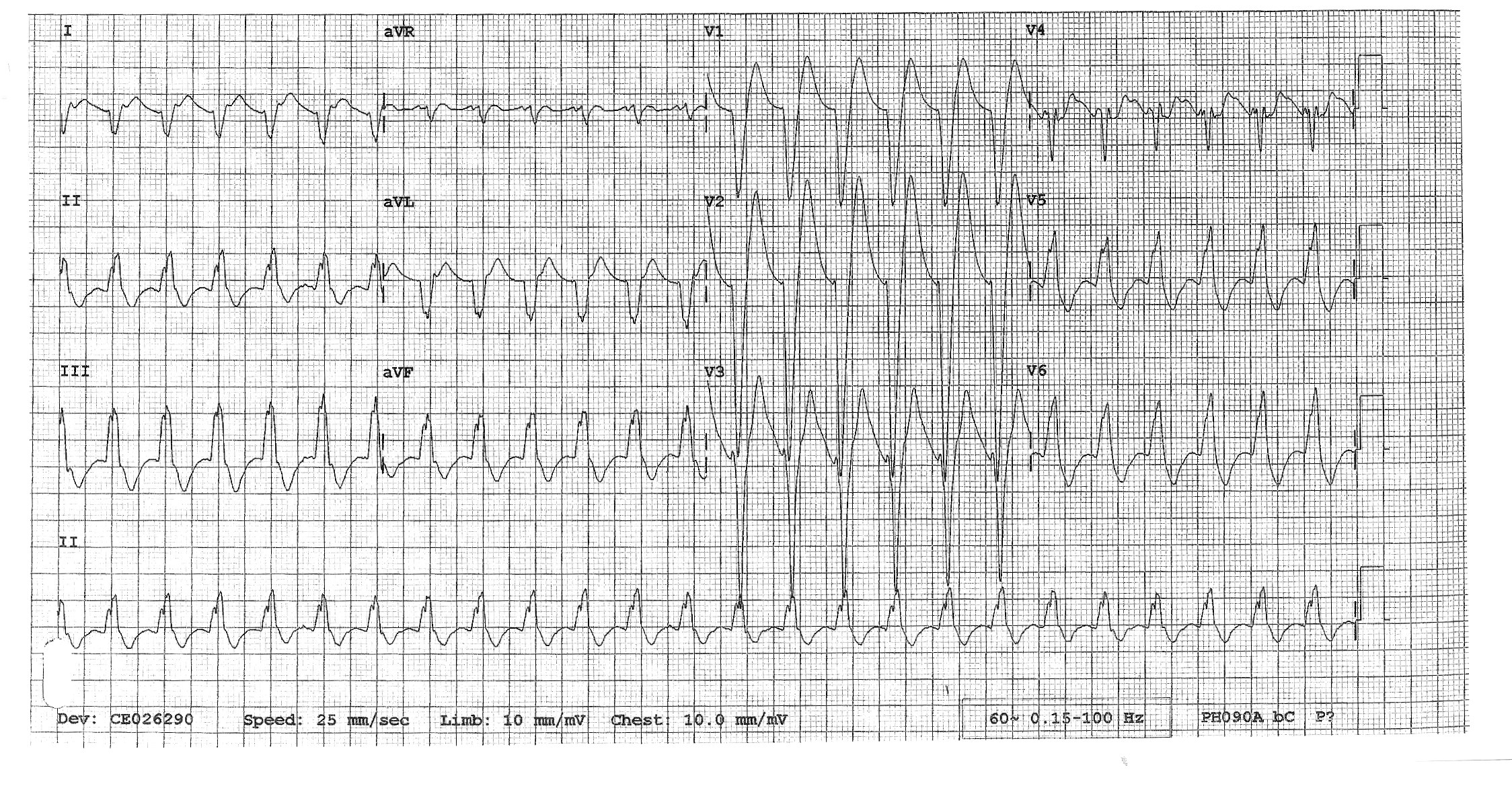
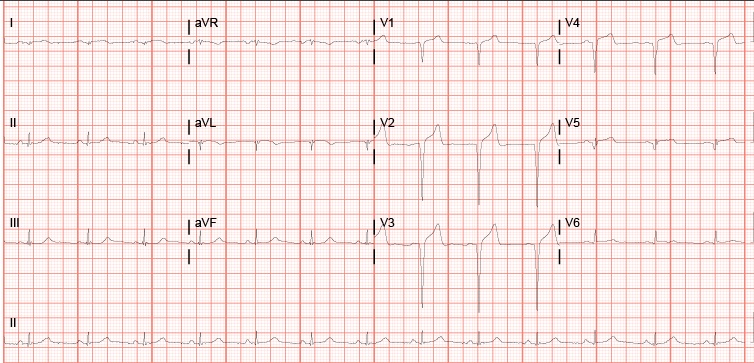
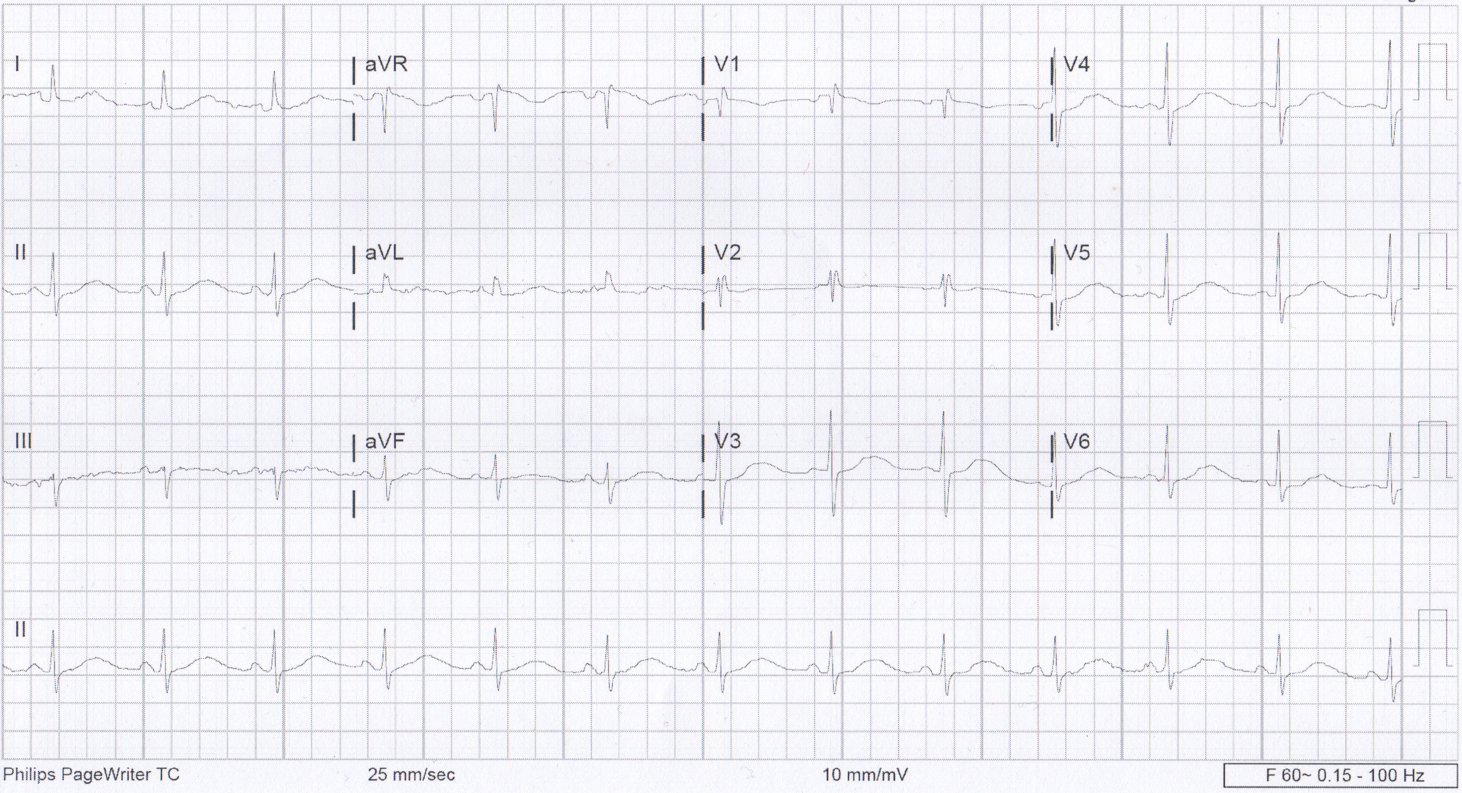
Pretty classic presentation here in a patient with previous heart disease.  His initial EKG recorded in triage is below:

Upon review, you can appreciate that there may be ST elevation in III.  Also, ST depressions are noted in the precordial leads along with ST changes elsewhere. My question to all of you based upon reviewing this EKG: Would you call this is a STEMI and would you activate the cath lab at this time?

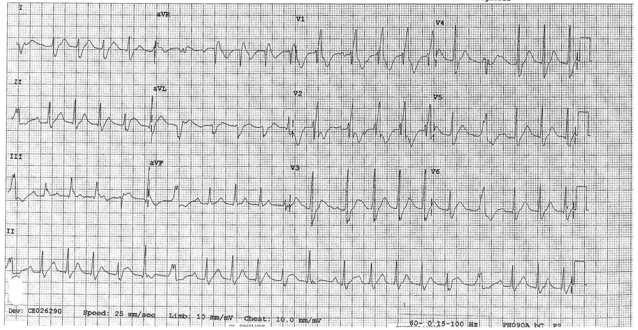
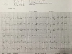
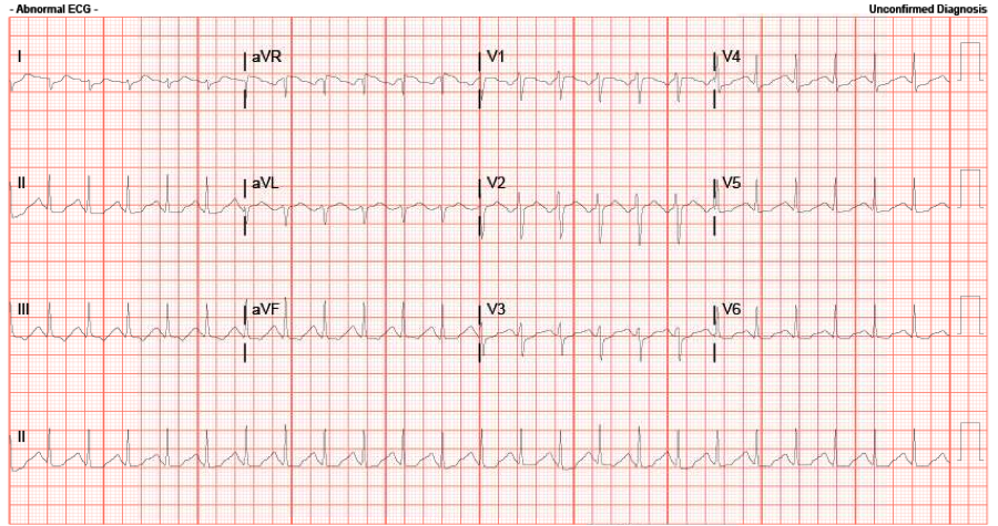
The attending that I was working with at the time didn’t feel that we could definitively call this a STEMI based upon the first EKG.  We examined the patient and collected his history after he was brought back.  After labs were collected, the patient received nitro paste.  Despite the paste, he continued to have pain. After approximately 15 minutes of being in the back, the patient reported that his pain was worsening.  The decision at that time was made to get a repeat EKG.  The repeat EKG is below:


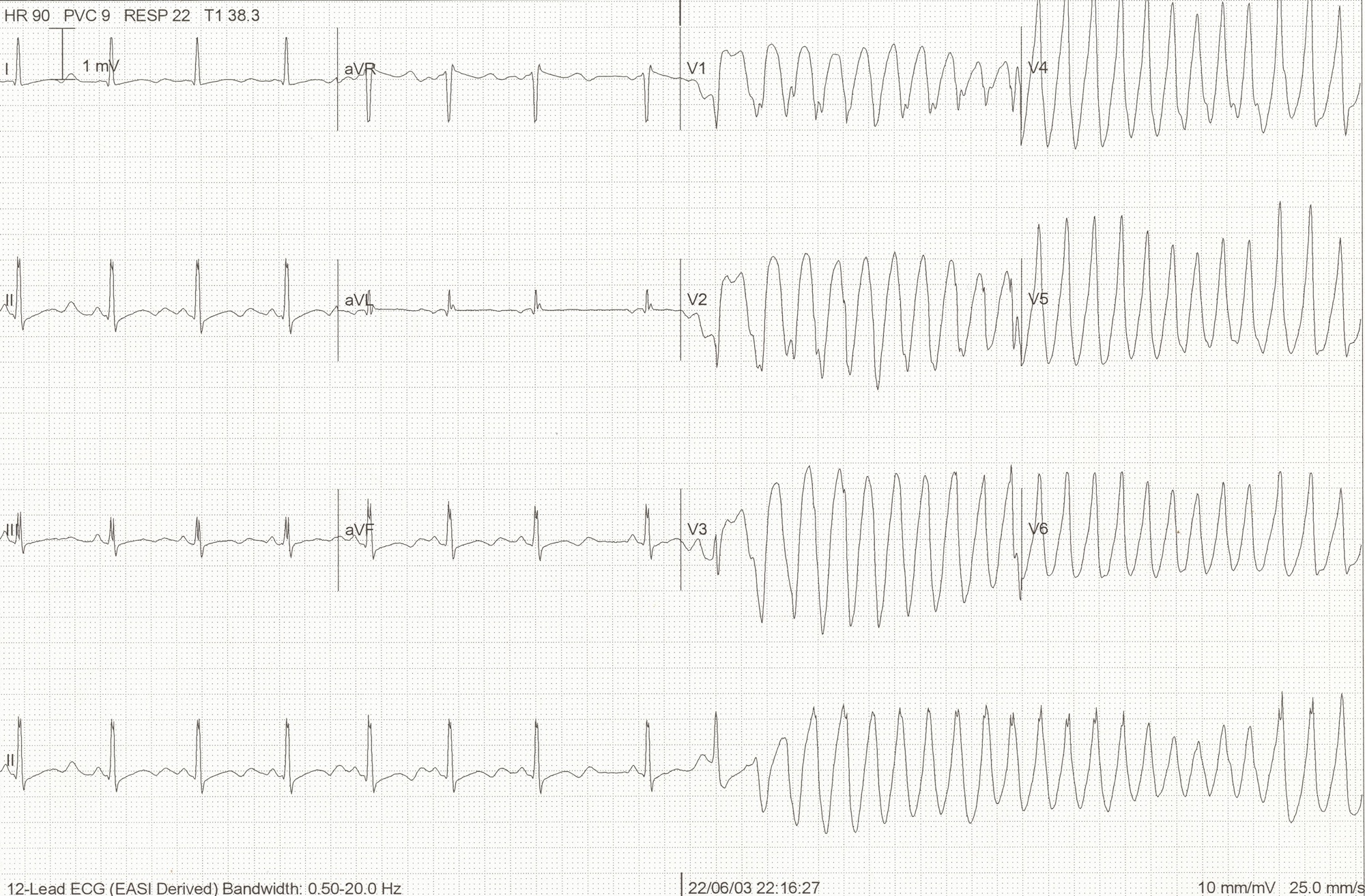
So now what are your thoughts? There is obvious ST elevation in the inferior leads.  There are also ST depressions in the precordial leads.  This EKG was taken 34 minutes after the first EKG which was performed in triage.  Through these two EKGs, you can appreciate the evolution of a STEMI.  This patient was emergently taken to the cath lab at that time and underwent left heart cath. He underwent an impella-assisted PCI to his SVG-PDA and was started on dual anti-platelet inhibitors. Following his PCI, he also had an episode of wide-complex rhythm which resolved after receiving amiodarone.

So this patient who was found to have a STEMI on his repeat EKG had presented with an initial EKG that was non-diagnostic for his condition. Did this affect his outcome in any measure? What can we learn from this?

According to a 2013 study conducted by Riley et. al, in a national sample of patients diagnosed as having STEMI (41,560 patients), 11.0% had an initial non-diagnostic ECG. Of those patients, 72.4% had a follow-up diagnostic ECG within 90 minutes of their initial ECG. There did not appear to be clinically meaningful differences in guidelines-based treatment or major inhospital outcomes between patients diagnosed as having STEMI on an initial ECG and those diagnosed on a follow-up ECG.

So, did the additional 30 minute delay in activating the cath lab affect this patient? Likely not. However, what this case reinforces is the importance of a repeat EKG as 11% of these patients likely will not have STEMI on their first EKG.


References:

Am Heart J. 2013 Jan;165(1):50-6. doi: 10.1016/j.ahj.2012.10.027. Epub 2012 Nov 21. Diagnostic time course, treatment, and in-hospital outcomes for patients with ST-segment elevation myocardial infarction presenting with nondiagnostic initial electrocardiogram: a report from the American Heart Association Mission: Lifeline program. Riley RF1, Newby LK, Don CW, Roe MT, Holmes DN, Gandhi SK, Kutcher MA, Herrington DM.

QTc

Yesterday I had a 21 yo female come in by BLS crew, tachycardic with a GCS of 3, spontaneously breathing with equal and reactive pupils bilaterally at ~ 6mm, with vomitus around her airway.  She was found down at home after her significant other called EMS due to concern that she was going to attempt suicide.  Report received that she was found with multiple empty pill bottles nearby.  No response to Narcan either in the field or in Room 9.

Here is her ECG: Calculated QTc (automated) is 401 ms, rate is 143.   The accepted normal value for QTc is: below 450ms for men and below 460ms for women.  The 99th percentile of normal: 470ms (men) and 480 ms (women).

ecg_case_micu

Just how does the computer calculate this?

By using the Bazett formula: QTc = QT / sqrt( R-R interval in seconds)

This means of course that if our rate is 60 BPM, then our R-R interval would be 1000ms, (or 1 second), and thus our QTc = QT/sqrt(1); and therefore in this situation QTc equals QT.

In our particular case: the R to R interval is 10.5 boxes (thus 420 ms, or in seconds: 0.420).  The QT was autocalculated at 260 ms, and when using the Bazett equation, this gives us a QTc of 401 ms.

What if you cannot rely on the computer calculated QT (which certainly can be inaccurate)? Then calculate the QT yourself by finding the tangential intersection of the T wave downslope with the ECG baseline, and measuring the intersection distance from the start of the QRS.  See the diagram below:

qt_picture

Using this measurement principal, and (in our case) using the lead V2 where the p wave and T wave are 180 degrees out of phase, we obtain a QT ranging from 8-9 boxes (320-360 ms).  When using the Bazett, this gives us a calculated QTc of 493-555. Of course qualitatively we can tell the QTc seems long as it exceeds half the R-R interval, quantitatively this is an increased QTc of 38% from the auto calculated, and is certainly in the significantly prolonged QTc range.

Follow-up: TCAs and benzos positive on her drug screen.  She was started on a bicarb drip in the ED (placed 3 amps of bicarb in a bag of D5); pH 7.34, lactic acid 1.2.  She is supposedly on Flexeril (similar in structure to TCA and will light up as TCAs on the drug screen), treated the same, however appears to be less cardiogenic in toxicity:J Emerg Med 1995;13(6):781-5.  Pt is intubated and stable currently.

Click here for the EMCrit on TCAs (overview below):

  1. Bicarb drip:  Goals: QRS duration <100, hemodynamically stable, Na ~150, pH ~7.5.  Sodium and bicarb don’t rise significantly in severe toxicity, her repeat showed no change in either.
  2. Magnesium: may help, though risk of Torsades is low as long as the patient remains tachycardic.
  3. Lidocaine: even though lidocaine is another Na-Channel Blocker, it will antagonize the effects of the TCA-like medications.
  4. Watch the electrolytes (decreases expected in both K+ and Ca+):  Lytes and ABG Q1H; (My pt’s Ca+ dropped from 9.8->8.5 over 4 hours).
  5. Intubation: hyperventilate to ensure no hypercapnia (want alkalosis).  Sedate with versed or propofol to raise seizure threshold.
  6. ECMO: If everything else fails.

Prolonged QT

58 yo F presents to the ED for cough, chest pain, and fatigue for 1wk. She has sharp, atypical sounding chest pain. But she is 58 and has risk factors: HTN, HLD, 0.5ppd smoker x48yrs. Better get an EKG.

Prolonged QT EKG

Initial EKG

Awesome, no STEMI. Done with EKG right? Hope you didn’t miss that really long QT interval.

First, how do we measure the QT?

QTc image
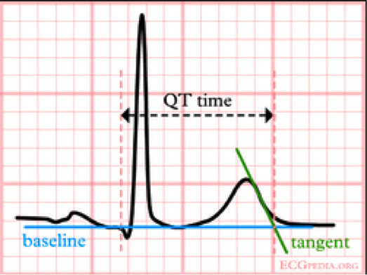
We usually talk about QTc rather than just the QT. This is because the QT interval varies depending on the HR. Using a correction equation standardizes the interval so it can be interpreted regardless of the HR. The EKG computer does give a calculation of the QTc, because we are obviously not hand calculating this on every patient. You should compare what the computer calculates to your gestalt when you review the EKG. If there are any concerns or discrepancies, you should hand calculate the QTc. MDCalc has an easy to use calculator. Above is the Bazett’s formula, which seems to be the most commonly used. Other formulas do exist. QTc is considered prolonged if > 440ms in men or > 460ms in women.

Next, why do we care?

ecg_hypokalaemia_torsades

This is the start of Polymorphic VT. There are several things that can cause this rhythm. Long QT is one possible cause. When Polymorphic VT is caused by a prolonged QT we give it a special name, torsades de pointes. The mechanism behind this is demonstrated on the above EKG. As the QT interval becomes more prolonged, there is a higher chance for an R-wave to hit on just the right part of the T-wave and cause this rhythm. QTc > 500ms seems be associated with higher risk.

So what causes prolonged QT?

Many things can cause prolonged QT. The most common etiologies are electrolyte abnormalities and drugs. Hypokalemia, hypomagnesemia, and hypocalcemia are well known to cause prolonged QT. Potassium and calcium are included on the CMP but don’t forget about magnesium. Drugs are also a big cause of prolonged QT. The list of drugs is long. Probably too long to memorize. However, there are some common medications and medication classes that you should know. The big classes are, Antiarrhythmics (like Amiodarone), Antihistimines (like Diphenhydramine), Macrolides (like Erythromycin), Antipsychotics (like Haloperidol), and TCAs (like Amitriptyline). This is not a complete list, just some highlights. If you really want to know if a specific medication is associated with prolonged QT, www.crediblemeds.org is a good source.

Other causes worth mentioning are structural heart disease, cardiac ischemia, and stroke. Some people do have Congenital Long QT Syndrome, but this should not be the leading diagnosis in the ED. Also those people are still at high risk for developing Polymorphic VT.

How did our patient do?

Her medications were reviewed and she was not on any of the most common offenders. Then routine labs came back. Unremarkable, except for K of 2.9! Repeat EKG after potassium repletion.

Normal EKG

EKG after K repletion


References:

  • www.uptodate.com
  • http://lifeinthefastlane.com/ecg-library/basics/qt_interval/
  • http://hqmeded-ecg.blogspot.com/2013/10/polymorphic-ventricular-tachycardia.html
  • bjsm.bmj.com/content/43/9/657/F3.large.jpgamp

Interesting case from the weekend – thoughts?

Hey guys, I was hoping to get your input on an interesting case I had at Kosair over the weekend.

16 yo F (6 ft, 150 lb…so basically an adult) with a PMH of depression, self injury, and prior suicide attempt presents after ingesting citalopram 40 mg x 90 pills (her prescription, just filled 2 days ago) and concerta 10 mg x 8-9 pills (her brother’s). Patient had been at a party the night before, admitted to EtOH.  Parents found out about the party the morning of admission and they had a big fight, took away car keys, etc. Patient decides to retaliate by swallowing pills, doesn’t tell anyone. Parents find her altered about 10:30, at Kosair at 11:40. Best guess is ingestion occurred sometime around 9-9:30am.  Had one seizure at home per family, and one en route per EMS. Generalized, tonic-clonic, brief.

Initial exam shows a drowsy but arousable patient. Answers orientation questions x3. Initial vitals show HR 147, BP 135/70, RR 25, 93% on some oxygen (can’t remember if NC or nonrebreather). Patient denies CP, palpitations, SOB, abd pain, N/V, weakness/numbness.  4mm, PERRL. MAE equally. Old self injury scars noted on wrists bilaterally. Exam otherwise unremarkable.

We start IVs, get her on a non-rebreather, get IV fluids going. Agree that charcoal seems like a bad idea with her mental status and seizures. Mom has shown up, and as we’re getting some additional history from her, respiratory is placing EKG leads. I’ve talked to poison control. Then, about 20 minutes into her stay, she seizes again. We bag her through the seizure, again generalized tonic-clonic, and just as we’re pushing 2 mg of IV Ativan she comes out of it. She appears post-ictal, but is maintaining her airway. We load her with Keppra, and as I glance at the monitor behind the attending’s head, I notice that her rhythm has changed and she looks like she’s got a wide QRS. We confirm she still has good pulses, still out of it mentally, and since she’s already connected to the EKG leads we grab one (time stamp 12:04):

EKG 1

By the time we get this printed off (!!!!) she appears to have spontaneously converted back to sinus on the monitor. But woah, holy wide QRS/long QT batman! As the attending and I are pouring over the first EKG we get another one immediately (time stamp 12:08):

EKG 2

Thankfully, the QRS appears to have normalized, but we’ve still got a loooong QT, one of the things poison control definitely told us to look out for. Having seen a few similar ingestions at University, I suggest it’s time for bicarb. The attending wants to confirm and we quickly call poison control back, they agree and suggest starting a bicarb gtt, with pH goal of 7.45-7.55.  Now we look back at the monitor and she’s throwing a ton of PVCs, captured here on EKG #3 (time stamp 12:12):

EKG 3

At this point, we opt to push an amp of bicarb while we’re waiting for pharmacy to tube up the bicarb gtt. I have to say, we see it start to work pretty darn quickly. The PVCs slow down, and her rate really starts to head back towards normal. We get an iStat (shot me down when I suggested one earlier), and a few minutes after we’ve pushed the bicarb we get an initial pH of 7.15, pCO2 of 51.5, HCO3 of 18, BE -11, and AG of 21. Electrolytes were WNL. By this time we also know her pregnancy is negative, and her serum tox is negative, no acetaminophen/salicylates on board.  At this point, we talk about intubation as the patient’s mental status is still waxing/waning and she’s breathing shallowly with brief periods of apnea, almost like an opiate overdose. Attending wants to hold off, so I go off to call the PICU resident…and end up having to hang up the conversation halfway through when he changes his mind.

So we intubate her (finally got a peds tube…in an adult), the bicarb gtt comes up from pharmacy, they’re cleaning the PICU bed her, the last EKG looks 1000x better, and all’s well that ends well (time stamp 13:08):

EKG 6

So I’m curious to see what your all’s suggestions/thoughts are on this case.  Looking back at that first EKG, how would you classify it? We’ve got a wide complex, monomorphic tachycardia that to me looks like sustained V tach (with a pulse).  The long QT doesn’t surprise me, but this rhythm does as you’d typically you’d worry about it devolving into Torsades, but that’s not what this is.

Looking back, things I would have done differently:  get a temperature sooner/order a total CK (serotonin syndrome could have been a factor and we don’t have a recorded temp until she’d almost 2 hours into her stay, no one ever ordered a CK), intubate sooner, loading her with keppra when she hit the door after 2 witnessed seizures, maybe could have prevented the 3rd?

Also, if you’re curious, I found this “Toxicology Conundrum” on LITFL that specifically discusses citalopram overdose. Has some good info, citalopram is definitely one of the more potent SSRIs, and QT prolongation is dose dependent and can be seen after ingesting >600 mg (this chick took 3.6 GRAMS). Seizures are also fairly rare, only seen in 2-3% of cases.

The elusive S1Q3T3

So this is a case I thought was interesting that I had in the department back in May. We all know that the most common EKG finding in the setting of PE is sinus tach, however the pimp question that is also asked is the finding of S1Q3T3. While I can easily recite the alphabetic-numeric code S1Q3T3 by heart at the drop of a hat, I had never seen one and honestly thought I never would. I thought finding S1Q3T3 was likely as rare as surviving a ED thoracotomy (OK maybe not that rare).  So on to the case.

39 year old female presents to the ED complaining of SOA and cough for the past two weeks. Cough was productive of green sputum, no fevers, and she does complain of some chest pain which sounds pleuritic in nature. Initial vitals HR 118 BP 111/73 RR 16 O2 97% room air. As I get into her PMH she says she has a history of multiple PEs with an IVC filter placed 1 yr ago because apparently she wasn’t very good at remembering to take her coumadin. She has had a hypercoagulability workup which was negative, no recent travel, no estrogen use.

So at this point with a history of multiple PEs, tachycardia, SOA, and pleuritic chest pain I am thinking I am going to scan this lady. Even though she had a IVC placed a year ago, she is still saying all the right things for PE. So while the CTPE protocol was cooking I got an EKG and there it was, S1Q3T3!

S1Q3T3

Needless to say I was pretty excited and immediately showed the rotating intern next to me who clearly didn’t share my enthusiasm. When I compared this new EKG to a past EKG a month ago she did not have the S1Q3T3.During her admission a month ago, when she had a normal EKG, she had a CTPE showing a chronic PE. This time when her CTPE came back the read was Acute on Chronic Pulmonary Embolism. So a month ago she had a chronic PE with a normal EKG and at this visit she had an EKG with S1Q3T3 and a acute on chronic PE. Out of curiosity I dug through her medical history a little bit more and found that this patient had multiple prior admissions for PE with multiple CTPE protocols and EKGs. What I found was that whenever this patient had a CT read of Chronic PE she had a normal sinus rhythm EKG. However, whenever she had a read of Acute on Chronic PE (which was 4 times!) she had a EKG showing S1Q3T3, dating all the way back to 2012. Yea, apparently this lady has been hanging out with a chronic PE in her distal right main pulmonary artery since 2012 and every once in a while she will throw a new small PE, even with an IVC filter.

So after doing a little bit of thinking and a little bit of reading it made sense. The EKG finding of S1Q3T3 is indicative of right heart strain, in this case resulting from an acute PE. So this patient’s heart has adapted to her chronic PE, however every time she throws a new PE she has an element of right heart strain which can be seen on her EKG as S1Q3T3.

I just thought this was pretty interesting to actually see the physiologic and mechanical adaption and strain this patient’s heart was undergoing being clearly demonstrated on her EKG. Also I learned that S1Q3T3 is not like a q wave after an MI in that it stays on the patient’s EKG, it is a finding that comes and goes depending on the patient’s presentation.

Anyways I thought this was a pretty cool little case and figured I would share. Hope you all enjoyed.

Reasons not to get into prison fights…

Middle aged male transferred from an OSH, accepted by ENT for a mandible fracture.

The patient is incarcerated, and was involved in an “altercation” with other inmates. The incident occurred around 2PM; but he didn’t report any of his pain to the guards until 10PM.  On arrival at the OSH he had multiple contusions to his face/head, lacerations over his hands, and obvious dental trauma.  The patient was also complaining of chest pain – he stated that another inmate had slammed him in the chest with his knee. Despite his age, the patient has a history of previous MI in 2011, cathed at U of L with no stents placed. Takes a baby aspirin, no other meds and no other PMH.

At this point, the patient is about 10 hours out from the incident. Work-up at the OSH with the following: neg CT head and CXR. CT face with a mandible fracture. Labs notable for WBC 17.8, Hgb 14.3, platelet 373, normal coags, normal electrolytes, BUN/Cr 14.0/1.1. Total CK 213 (55-170 normal), troponin <0.012, CKMB  1.66 (0 – 3.38 normal), myoglobin 271.8 (0-121 normal).  Tox screen negative. EKG is as follows:

OSH EKG

His hand lacerations were repaired and he was started on Augmentin for a human bite. ENT accepted, and the patient was transferred to U of L, arriving about 6 AM. Dental was consulted on arrival and splinted his teeth. By 9 AM ENT had evaluated the patient and admitted him to the floor, planning for surgical intervention.

The patient was an ED floor hold, and around 2PM began complaining of worsening chest pain. ENT was paged and ordered an EKG and a set of cardiac enzymes, coming down to re-eval the patient. His EKG now looked like this:

1410 EKG

Enzymes came back with CK total 5024, CKMB 303, and troponin 44.1. Cardiology was consulted and ordered a stat echo and started the patient on ACS protocol. The echo showed an EF of 30%, an akinetic mid/distal anferoseptum and an akinetic apex. Cards initially thought that this was consistent with stress cardiomyopathy in the setting of trauma, but couldn’t rule out cardiac ischemia due to direct cardiac trauma. They planned to treat medically and cath in the morning.

Throughout the evening, he developed worsening ST elevation in his lateral leads and his troponin continued to rise, up to 67.0 by midnight.

0308 EKG

The on call cath attending at Jewish was consulted and by about 3AM the decision was made to transfer the patient to Jewish for a cath first thing in the morning.

Final result: 100% LAD occlusion, secondary to direct cardiac trauma.

Definitely rare injury, but one to keep in the back of your mind, especially as it can occur in previously healthy, relatively young patients. Of note, these can have delayed presentations, up to several days. Typically occur after MVA, but there are several cases reports occurring after crush injuries, being hit in the chest by a soccer/rugby ball, and my personal favorite, one listed as “struck in the chest by an umbrella tip.”

Death of CK-MB?

Hey guys here is a relevant (albeit 7 years old) article supporting my abandonment of the CK-MB. I had heard Mattu talk about this on some podcasts but this article (along with some newer ones) is the evidence to support that practice. At Jewish I do not obtain a CKMB though when I want a total CK I now have to remember up front. On the POC machines at Jewish the Trop alone seems to error far less than when CKs are running with it. Still watch out for the Trop false positives.

Also I would plug Dr Mattu’s new ECG site. Now he charges a small fee (can pay weekly or monthly but I did the $27/year). Still video based (I prefer to just read a blog with pics) but top notch quality. There is a weekly case and a monthly lit review. The above article is in his Feb review. Might make a one month subscription part of the ECG month. ECGs of course are learned over years and decades, not in a month long elective.