QTc

Yesterday I had a 21 yo female come in by BLS crew, tachycardic with a GCS of 3, spontaneously breathing with equal and reactive pupils bilaterally at ~ 6mm, with vomitus around her airway.  She was found down at home after her significant other called EMS due to concern that she was going to attempt suicide.  Report received that she was found with multiple empty pill bottles nearby.  No response to Narcan either in the field or in Room 9.

Here is her ECG: Calculated QTc (automated) is 401 ms, rate is 143.   The accepted normal value for QTc is: below 450ms for men and below 460ms for women.  The 99th percentile of normal: 470ms (men) and 480 ms (women).

ecg_case_micu

Just how does the computer calculate this?

By using the Bazett formula: QTc = QT / sqrt( R-R interval in seconds)

This means of course that if our rate is 60 BPM, then our R-R interval would be 1000ms, (or 1 second), and thus our QTc = QT/sqrt(1); and therefore in this situation QTc equals QT.

In our particular case: the R to R interval is 10.5 boxes (thus 420 ms, or in seconds: 0.420).  The QT was autocalculated at 260 ms, and when using the Bazett equation, this gives us a QTc of 401 ms.

What if you cannot rely on the computer calculated QT (which certainly can be inaccurate)? Then calculate the QT yourself by finding the tangential intersection of the T wave downslope with the ECG baseline, and measuring the intersection distance from the start of the QRS.  See the diagram below:

qt_picture

Using this measurement principal, and (in our case) using the lead V2 where the p wave and T wave are 180 degrees out of phase, we obtain a QT ranging from 8-9 boxes (320-360 ms).  When using the Bazett, this gives us a calculated QTc of 493-555. Of course qualitatively we can tell the QTc seems long as it exceeds half the R-R interval, quantitatively this is an increased QTc of 38% from the auto calculated, and is certainly in the significantly prolonged QTc range.

Follow-up: TCAs and benzos positive on her drug screen.  She was started on a bicarb drip in the ED (placed 3 amps of bicarb in a bag of D5); pH 7.34, lactic acid 1.2.  She is supposedly on Flexeril (similar in structure to TCA and will light up as TCAs on the drug screen), treated the same, however appears to be less cardiogenic in toxicity:J Emerg Med 1995;13(6):781-5.  Pt is intubated and stable currently.

Click here for the EMCrit on TCAs (overview below):

  1. Bicarb drip:  Goals: QRS duration <100, hemodynamically stable, Na ~150, pH ~7.5.  Sodium and bicarb don’t rise significantly in severe toxicity, her repeat showed no change in either.
  2. Magnesium: may help, though risk of Torsades is low as long as the patient remains tachycardic.
  3. Lidocaine: even though lidocaine is another Na-Channel Blocker, it will antagonize the effects of the TCA-like medications.
  4. Watch the electrolytes (decreases expected in both K+ and Ca+):  Lytes and ABG Q1H; (My pt’s Ca+ dropped from 9.8->8.5 over 4 hours).
  5. Intubation: hyperventilate to ensure no hypercapnia (want alkalosis).  Sedate with versed or propofol to raise seizure threshold.
  6. ECMO: If everything else fails.

Name that Disease?

55 y/o  F with hx of HTN,COPD and recurrent indurative lesion on her left foot, last event was a yr or so ago, presented to the ED for worsening pain and increased size of her lesion for the last few wks now. No fever, chills, or fatigue. On exam, the cutaneous lesion is mildly tender and erythematous, non-fluctuant, no warmth noted. Pt is immunocompetent.

img_7436


/

/

/

/

/

/

/

/

/

/

/

/

Answer:

Pyoderma Gangrenosum.  Take home point is to not I&D this lesion. It is not an abscess. It is a rare autoimmune disease that affects pts in their 40s-50s. These pts will have hx of other autoimmune diseases–lupus, crohns etc.  An I&D would lead to phenomenon known as pathergy,  the formation of new lesions following a trauma.

Tx: High dose steroids and pain meds. Refer to podiatry. Pt in this case was already well known to podiatry on arrival and was discharged with steroids and pain meds after podiatry consult in the ED.

Approach to the Fussy Infant

There are many different types of challenging patients that we all dread seeing when they pop up on the board. Whether the patient’s chief complaint is headache, back pain, or pregnant female with abdominal pain. Another very challenging patient presentation is the crying infant. The differential when evaluating a crying infant is broad. In this post I will include a list of differential diagnosis to consider based on organ system and then a patient I had that presented in this way.

CNS- Meningitis, epidural hematoma, subdural hematoma, hydrocephalus

HEENT- Skull fracture (accidental or non- accidental trauma), ocular foreign body, corneal abrasion, otitis media, nasal foreign body.

Cardiovascular- SVT, myocarditis, congestive heart failure

Pulmonary- foreign body in airway, bronchiolitis, pneumonia

GI- Malrotation/volvulus, pyloric stenosis, appendicitis, gastro-esophageal reflux, intussusceptions, anal fissure

GU- Testicular torsion, UTI, incarcerated inguinal hernia, soap vaginitis, phimosis, paraphimosis

Musculoskeletal- Fracture, septic arthritis, dislocation, hair tourniquet

My patient presented as a 1 month old male with his Spanish speaking Hispanic parents. Mom stated that he has been crying consistently for the past 4 days. She does not remember a specific time when the crying started but she states it has not improved.

Mom is breastfeeding the baby and denies any dietary changes. The baby was full term and mom had no complications with the pregnancy. Patient up to date on all vaccinations. Baby has been afebrile and per mom has not been lethargic. Baby is still feeding well and gaining weight appropriately and mom denies any projectile vomiting after feeds, denies any change in stooling, and notes good urine output. Baby lives at home with mom and dad and I have no red flags to suspect non-accidental trauma.

On exam he is overall well-appearing and does not appear to be in any type of distress: crying but consolable. He appeared to be healthy. He was interactive, tracking me with his eye movements, and did not appear to be meningitic or lethargic. Heart and lungs were unremarkable and abdomen was soft, nontender and non distended. GU exam (make sure you do this) was unremarkable. Baby was moving all extremities while lying on the oversized adult bed in the middle of the hallway and on inital exam did not appear to have any outward bruising or signs of trauma.

Upon removing the patient’s socks I noticed something odd on his second toe of his right foot. Just distal to his PIP joint he had a circumferential red line. As soon as I started to examine that toe his crying increased substantially. On further examination he had a hair tourniquet that had eroded its way all the way down to the bone of the middle phalanx of the second toe.

At this point the baby was still in the hallway and we took him to Room 9 to attempt to try any remove it. I am sure you can imagine how awesome this was on a crying kicking 1 month old. We attempted to unwind the hair but ultimately were unable to do so as it was just to deep into the tissue. I called Kosair and the patient was transferred and I do not yet follow up on the final outcome.

This just re-enforces the importance of a good head to ahem, toe physical exam on patient’s that are not straight forward. Mom had been with the infant 24/7 for the past 4 days and had not noticed this; not to mention who knows when the hair tourniquet actually started. Just something to keep in mind and hopefully this helps next time you all have to examine a crying baby.


Central Line Insertion Choice

All,
I know this came up during conference today so thought I’d send the article I think was cited. At least this is the one I found from EMRAP, below is their summary of this article. Long and short of it, complication rates are really low, when done in sterile fashion in a controlled environment.
This was done in the ICU, not in the emergency department.
Overall they didn’t say that one site was absolutely the best.

nejm-2015-central-line-site-complications

Take Home Points
No central line site is superior.
Femoral lines are fastest and most successful. Subclavian lines have a lower infection risk but higher rate of pneumothorax.

Parienti, JJ et al. Intravascular complications of central venous catheterization by insertion site. N Engl J Med. 2015 Sep 24;373(13):1220-9. PMID: 26398070

Bottom line: no line was superior. Femoral lines were the fastest to place and had the highest success rates. Subclavian lines had the lowest infection risk but had a higher rate of pneumothorax.

A patient needs central line access. Which should we choose? Which is best? There are multiple complications; infection, mechanical complications like artery puncture or pneumothorax and thrombotic complications.

The authors of this study conducted a randomized, controlled trial in 10 French ICUs. They enrolled adult patients with at least two accessible sites. Patients with all three sites accessible were randomized in a 1:1:1 fashion while those with only two sites were randomized in a 1:1 fashion. The doctors had all performed at least fifty central lines. However, they were all aware of the study and probably tried harder to reduce complications.

They looked for symptomatic clots and/or infection from the time of insertion up to 48 hours after removal. This was a large study; 3471 catheters were placed in 3027 patients. Catheters were assigned to a randomly assigned site and side; placement was successful approximately 91% of the time. 85% of subclavian lines were successfully placed, 91% of the jugular lines were placed and 95% of the femoral lines were placed. Femoral lines were most successful and subclavian lines were least likely successful.

Placement of femoral lines was also more rapid, by about a minute.

The primary outcome was a composite of infection, symptomatic clot and mechanical complications such as pneumothorax and bladder puncture. The jugular line performed the worst followed by the femoral line, then subclavian line. However, it is important to look at the individual components.
For mechanical complications, the subclavian line performed the worse. 2% had a complication versus 1.5% of jugular lines and less than 1% of femoral lines.
All lines were fairly low for symptomatic clots; 0.5% for subclavian, 1% for jugular and 1.4% for the femoral group.
In terms of infection, the subclavian group was the lowest (0.5%). The highest? Surprisingly, jugular lines had a 1.4% rate of infection versus 1.2% in the femoral line group.
Overall, there were fairly low rates of complications. These were performed in very sterile conditions in the ICU. These were not placements in crashing or coding ED patients.

Placement of central lines, including femoral lines, when done carefully under sterile conditions has a low rate of complications. This article does not identify one superior line placement.

Headache in a post-neurosurgical patient

Earlier this month while moonlighting I had an approximately 40yM present for a headache (9/10 pain) for 3 days.

I know, this is an everyday occurrence.

But in his case he had a craniotomy with removal of a meningioma 1 month prior. He also noted 2 weeks of swelling on the left side of his head along the surgical scar.
ROS: denied fevers, chills, changes in vision, weakness, numbness, or tingling, etc.

PE: VSS, HEENT: Left side of head extending from his surgical scar and wrapping around to even under his L eyebrow was swollen and firm. NEURO: WNL

So, I know something isn’t right and my guess is that he either had a bleed or infection associated with his surgery. I order a CT head. For his headache I give him a migraine cocktail (IVF, compazine, and benadryl, minus the toradol).

img_1534

img_1535

After I see the CT images, I rush to check on the patient as the CT obviously shows quite a bit of midline shift and the patient states his headache is drastically improved (2/10 down from 9/10). Apparently migraine cocktails work on all causes of headache.

I consulted Neurosurgery who promptly admitted the patient to the ICU with plans to go to the OR. Approximately 45 minutes later, after the NES nurse practioner has seen the patient and he is getting packed up to go to the ICU, the radiologist calls to notify me of the CT findings.

This is a reminder to ALWAYS look at CT images yourself, especially if you’re expecting a life threatening finding.

Chest pain?

Yesterday, 10 minutes before the end of a very busy shift in the middle of nowhere:
Nurse hand’s me an EKG for the mom of one of our best young medics. Ischemic STD in II, III, aVF, V5, V6. No STE. Maybe some LVH. No other EKG on file, she hasn’t been here before. I get up to see her right away.
In the room is a 50 yo lady in severe distress. BP 250/140. Describes “tearing” L sided CP radiating to her upper back. I ask her if she has a family hx of sudden death or aneurysms, her son says yes. No SOA. No N/V. Lungs are clear. Can’t palpate pulses in her feet. Slightly obese. She is sweating. There isn’t a CT surgeon in this county; I am sweating, too.
The nurse gets CT ready and I start the ball rolling for big IVs, blood, helicopter, etc. She gets dilaudid 1 mg then 0.5 then 0.5 again for pain and hopefully BP control. I tell everyone she’s now the ED’s top priority. The family of another patient grabs me twice in the hallway and asks why their mom, who fell, hasn’t gotten her home dose of lisinopril yet.
~30 minutes from door to imaging – CTAs = no dissection. Great, I have some time. Pain is well controlled now and BP 150s/80s. Repeat EKG w ischemia resolved completely, normal. Hypertensive emergency? She has a bit of a headache so I scan her head because we’re in CT, but it’s not useful due to the residual contrast. Trops, CBC, CMP, urine, tox, etc. all negative. Now looks great and feeling much better. Wants to go home.
Her son, her nurse, and I spend a long time convincing her she needs to stay overnight. She doesn’t want to be admitted, as she’s starting a new job Tuesday and can’t miss it. Eventually, she agrees to stay. Then we have to convince her to be transferred, since ED MD is the only MD in house overnight and she had me straight terrified. Reluctantly, she agrees to the transfer as long as her son will bring her home Monday night, no matter what. Hospitalist at the local mothership accepts readily. At that hospital, she’d had a negative stress 8 months ago and NES had placed her shunt (pseudotumor, she has a lot of headaches); hospitalist will consult cards and NES when she gets in.
I arrive back to work this morning to find out she’d made it upstate only to be transferred again overnight. She’d been flown to a bigger center after repeat CT head demonstrated SAH. She had 2 aneurysms, 1 was bleeding, both were definitively addressed. 24 hours after walking in the door, at the time of this writing, she is extubated and thriving.
Found this article from 1988 with a similar presentation, but I can’t say I’d ever heard of it before. Something else for your differential. Really consider that admission for observation when something seems wrong, even if you don’t know what it is.
Going to change my drawers now.

Delivery in the Emergency Department

I will apologize for the wall of text in advance but I thought I to share an experience from last May that fits pretty well with the first 2 weeks of lectures this month. I think it definitely highlights the importance of  feeling comfortable with both delivery and newborn resuscitation in case they actually happen to you. I’ve also tried to point out all problems that came about but I am sure I didn’t hit them all and I am positive that I could have handled some of them better. 

It was nearly the end of a pretty typical shift at Jewish Downtown. For those of you who haven’t been there or don’t know there is zero OB coverage at Jewish. I was finishing my charts when I hear the secretary say “pregnant woman in labor” over the phone. That is a pretty unusual thing to hear at Jewish so I immediately asked the attending who was sitting closer if I heard that correctly. I also half-jokingly said I definitely wanted in on the patient if they were serious because I still needed a few more deliveries. The attending chuckles and replied that she thought the secretary was only joking because there shouldn’t be any reason for a laboring patient to come to Jewish, so I returned to my charts.

About 2 minutes later they call for a physician from the room behind the doctors area and we walk into what is in fact a laboring patient. The nurses state she seems to be contracting every 3-4 minutes but they don’t know much else at this point. While the attending checks her cervix, I grab the ultrasound and check the fetal position. She was about 8cm dilated and the best I could tell the baby was vertex but the head was so far down I gather this from the rest of the anatomy. Immediately calls start going out to the neonatologist at Kosair while we attempt to talk to the family and figure out how the patient ended up at Jewish. Here comes out first problem:

Problem 1: Neither the patient nor the family speak a word of english. Not only that but there is no translator phone in the room so the family has to be taken to another room to try to get some questions answered.

In the meantime I attempt to get a fetal heart rate to assess how the fetus is doing.

Problem 2: The only doppler is a pen style for checking pulses that doesn’t actually display a pulse number.

I go back to the ultrasound, find the heart and count the beats on the screen while a nurse counts for 15 seconds to get a FHR of about 144, which is always reassuring. In the mean time we are able to obtain some more information from the family.  

Apparently, the patient’s water broke during her office visit around 2pm (it’s now almost 11pm) and she was told to go to the hospital. For whatever reason they decided to wait and they got mixed up between Norton’s and Jewish hence our current situation. There are calls being made to Norton’s L&D informing them of the situation and transport is on the way. The neonatologist is also en route as a safety precaution in case she actually delivers here. The patient and fetus appear to be stable, and while still contracting around every 3-4 minutes she still isn’t fully dilated. I make the mistake of leaving the room assuming that the patient will soon be swept away to have her baby properly on an L&D floor. About 5 minutes later another call from the room and I walk in to see the beginning of this baby crowning.

The nurses wheel in their delivery kit as I gown and glove up. I apply a few packets of lubricant jelly, have the nurses move the patient closer to the end of the bed and attempt to create a semi-sterile field with the supplies available.

Problem 3: It’s nice and easy to set up to deliver in a room with a bed designed to deliver a baby. Unfortunately for me this situation involved a nurse/tech on each leg and the patient mostly laying flat in a bed with bag to collect fluid half hanging off the table but mostly just shoved under her bottom as best as I can with no real drainage.

Problems 4 & 5: Did I mention she didn’t speak english, also I have no idea what the word for “push” is in her language.  Also when you are on the L&D floor you have all kinds of cool toys such a tocometer to help you know when to tell the patient to push. I do not have that luxury.

I attempt to put a hand on her abdomen to feel her uterus contract so I know when to tell her to push and hope that she figures out what we want from her. Maybe it was a good thought but I have no idea, good thing she does and she is pushing every few minutes and the head is progressing it’s way out.. After a few good pushes the progress seems to slow a little bit and I start to worry a little bit that this 2 week post-dates baby may be stuck. What was that mnemonic for shoulder dystocia again? All I can remember is McRobert’s maneuver, but for anyone curious, Rosen’s has a nice one:

Help: Obstetrics, neonatology, anesthesia

Episiotomy: Generous, possibly even episioproctotomy

Legs flexed: McRoberts’ maneuver

Pressure Suprapubic pressure: shoulder pressure

Enter the vagina: Rubin’s maneuver or Wood’s maneuver

Remove posterior arm Splint, sweep, grasp, and pull to extension

Luckily, I have the nurses holding onto each leg (because this bed doesn’t have stirrups) so they flex her legs towards her as much as they can and everything continues to progress smoothly. Eventually the head is out and the rest of this baby boy delivers quickly. He is suctioned, wrapped in a warm blanket and the nurses begin assessing him. I think we put his APGAR at a 7 at 1 minute.

So I am done right? Baby is out, nothing more to see or do, lets ship them out. Right?……

Turning back to the patient I realize she seems still be bleeding a little more than I expected. It’s hard to figure out where she is bleeding from so I deliver the placenta which appears to be intact. I even sweep and massage the uterus just to be sure which seems to be contracting well.

Problem 6(?): Not really a problem because it seems her bleeding was not coming from the uterus but I have no access to the medications typically used to help control uterine bleeding after a deliver such a pitocin. Not that it matters because I didn’t know what the dose would be anyway. Just another interesting thought that I had during this whole process.

Since the placenta is whole, and seems to be firm I look for other sources of bleeding. This is when I realize the patient has a nice 2nd or 3rd degree tear (Dr Sterrett would be very disappointed I didn’t control the head well enough). I check to make sure it isn’t a 4th (thankfully it’s not), and start contemplating my next course of action. The attending asks what kind of suture I want to use to repair her tear. My only reaction was to smile and say “nothing”, followed by explaining that since the bleeding is slowing it’d probably be better to let the OB-GYNs fix her. I plan to pack her to make sure she doesn’t bleed too much in transport. 

Problem 7: No one had even seen a vaginal packing kit in the ED before, so we improvised and used some kerlex with a tail for easy removal.
By the time this had finished the neonatologist had arrived and began assessing the newborn. I think we ended up giving him apgars of 7 and 9. I started the patient on some fluids (because we didn’t have an IV when all of this started, another mistake I didn’t realize until it was all over) and within another 2 minutes transport had arrived to take the patient to Norton’s and she was actually swept away. Hopefully the OBs she finally got to didn’t think I botch the whole thing too much. Overall it was a pretty intimidating and adrenaline pumping situation. Not sure if I will ever have an experience quite like this again but if it does happen at least it will not be the first. 

Any comments, critiques, criticism or otherwise are welcome.

1.4% Observed Adverse Reaction Treated With Flumazenil

Flumazenil (Rx: Romazicon) has recently been described as coming into favor for two unique purposes: (1) hepatic encephalopathy and (2) paradoxical reactions to benzodiazepines.

Regarding the first, flumazenil’s use in hepatic encephalopathy has been well described recently in a Cochrane review of 113 RCTs with a total n = 805, wherein flumazenil had a significant beneficial effect on short term improvement of hepatic encephalopathy.1 This is thought to occur physiologically secondary to reversal of the origin of hepatic encephalopathy—i.e., an accumulation of substances that bind to a receptor-complex in the brain resulting in neural inhibition1 (principally GABA receptors which are forefront in the stimulation of sedation). Therefore GABA receptor antagonists (such as flumazenil) can be used to directly oppose this mechanism. Effect on full recovery and survival has still not been proven with flumazenil administration.1

Secondly, flumazenil can be used for paradoxical reactions to benzodiazepines2,4 and in a 10 year review of its use, published in the Journal of Emergency Medicine,3 the real safety of this drug has once again come into question, as there were relatively few adverse outcomes even in the highest of seizure provocation risk—which occurred with co administration of pro-convulsant (e.g., TCAs) at a 2.7 % incidence (8/293)—the total incidence including all subjects bore a rate of 1.4% of seizure activity (n = 904).3

I present an example of administration in the second of indications above. I took care of a 26 yo WF with PMH of asthma, a prior severe dental cavity pending root canal and an IV heroin addiction, currently sober and progressing through the the 12 Steps program at the Healing Place. She presented in sepsis, afebrile with qSOFA of 0/3 (Labs: WBC 21.2 with left shift, procal 1.33, ESR 83, CRP 201, lactic acid 0.8 s/p 2 L NS IVFs), and AKI (Cr. 1.6) with dental as well as urinary possible sources. She was eventually discharged on day 3 with Dx of urosepsis, creatinine returned to normal, and had a negative echo for routine endocarditis rule out in the setting of PMH of IVDA.

During her ER stay she was uncomfortable, diaphoretic, pale, GCS of 15, but anxious and in pain, professing severe insomnia for 3 days, stating, “I just want to sleep”. A trial of oral Ativan 2 mg was given, as she did not want any pain medication due to her prior addiction. She noted a small temporary improvement; however 2 hours later this beneficial effect was absent. By now she had received cefepime 2g and vancomycin 25 mg/kg (for potential osteomyelitis coverage), and was requesting more anxiety medications, having already received 50 mg IV Benadryl 30 minutes prior with no improvement noted. Clinically she was GCS 15, pleasant in interaction, increasingly pale, uncomfortable, wide awake at 0445, and subjectively in pain. She was then given 2 mg IV Versed.

Immediately following the administration of midazolam she became altered to GCS 12 (E4, V3, M5), eyes wide, extremities tremulous, pulled out all of her IVs, and was trying to jump off the bed. It was clear she was paradoxically agitated and hyper-aroused. Rather than reversing her (though we doubted history of benzodiazepine use), we opted to watch and see if this reaction would subside without intervention since she responded favorably to the oral Ativan; however the rarely seen but well known paradoxical reaction to Versed was suspected. She was observed 1:1 and thereafter 3:1 for 40 minutes, at which time she appeared to be steadily worsening rather than improving. The decision was made to give an IV push of 0.2 mg of flumazenil (Rx: Romazicon). Within 30 seconds after administration she once again returned to her pleasant self, she was GCS 15, appropriate, and had no recollection of the previous hour, and had no seizure activity noted throughout her stay. She maintained a healthy mental status of GCS 15 and was AAOx4 for the rest of her evaluation and admission.

In 2010, Kreshak et al. reported a similar case and treatment. This paradoxical reaction to Versed in their report is thought to occur at less than 1% incidence, however it is described as commonly as 1.4 %.4 In the reported literature this reaction is described as a patient becoming acutely agitated, restless and aggressive2. Stiffening and jerking of the extremities, and shaking of a part of the body are also noted. When observing a patient with this reaction, after ruling out other etiologies of agitated AMS, Kreshak et al. (2010) opted to administer flumazenil 0.5mg IV, and “…immediately after which the patient became conscious, oriented and calm, the paradoxical reaction was terminated”. The patient had no recollection of the events,2 similar to the patient observed in the ULED.

Per Kreshak et al. (2010), there exist “…different theories concerning the mechanism of paradoxical reactions, involving a central cholinergic effect or the serotonin imbalance”.2 Unfortunately the exact mechanism of paradoxical reactions remains unclear.

Although difficult to locate literature, if seizures develop following flumazenil administration, pharmacology guidelines recommend Valium 20-30 mg IV then immediately switching to barbiturates; some soft EM sources also suggest going straight to propofol.5

Thank you for reading my post.

References

  1. Als-Nielsen, B., Kjaergard, L., & Gluud, C. (2001). Benzodiazepine receptor antagonists for acute and chronic hepatic encephalopathy. The Cochrane Database of Systematic Reviews (Complete Reviews). doi:10.1002/14651858.cd002798
  2. Cabrera, L., Santana, A., Robaina, P., & Palacios, M. (2010). Paradoxical reaction to midazolam reversed with flumazenil. Journal of Emergencies, Trauma, and Shock J Emerg Trauma Shock, 3(3), 307. doi:10.4103/0974-2700.66551
  3. Kreshak, A. A., Cantrell, F. L., Clark, R. F., & Tomaszewski, C. A. (2012). A Poison Center’s Ten-year Experience with Flumazenil Administration to Acutely Poisoned Adults. The Journal of Emergency Medicine, 43(4), 677-682. doi:10.1016/j.jemermed.2012.01.059
  4. Tae, C. H., Kang, K. J., Min, B., Ahn, J. H., Kim, S., Lee, J. H., . . . Kim, J. J. (2014). Paradoxical reaction to midazolam in patients undergoing endoscopy under sedation: Incidence, risk factors and the effect of flumazenil. Digestive and Liver Disease, 46(8), 710-715. doi:10.1016/j.dld.2014.04.007
  5. (n.d.). Retrieved August 23, 2016, from http://www.goodfriendem.com/2013/05/flumazenil-romazicon-is-probably-safer.html

5 day old with “seizures”

Recently I had an interesting case at Kosair of a 5 day old male who presented with jerking movements of his arms and legs. He always had “twitches,” which the parents had been assured were normal for a newborn, but the episodes were getting worse. Since the day before, he had had several episodes where both arms would shake and seize up and his legs would curl up under him, lasting about a minute. He is sleepy afterwards, but mom thinks he’s always pretty drowsy. Overall it was unclear if what mom was describing was a seizure. Even her helpful phone videos were not 100% clear, but we proceeded as though they were real seizures. In a 5 day old.

Mom had 3 UTIs during pregnancy, and her labor was likely precipitated by an episode of pyelo. She and baby were briefly tachy during labor but pain meds helped, and the SVD was otherwise uneventful. No STIs, GBS negative.

Baby was afebrile, normal VS. Appeared drowsy until the usual screeching during the cath urine, so overall, well-appearing baby. He did twitch sometimes, but he never had one of the spells while he was in the ER.

Differential diagnosis for neonatal seizures? Bacterial meningitis, viral encephalitis, intraventricular hemorrhage, SAH, SDH, hypoxia, hypoglycemia, hyponatremia, inborn errors of metabolism, etc.

Our patient wasn’t actively seizing and labs were WNL.

Subdural hematoma (from birth) and meningitis were high on our differential. We went ahead and gave antibiotics but got a CT head before proceeding with the LP, and I’m glad we did.  It turned out that he did have a subdural hematoma, likely parturitional. We elected to forego the LP, since he was afebrile and we already had a reasonable explanation for his symptoms. Neurosurgery wanted a repeat CT in 6 hours (surprise!), and neuro wanted an EEG (surprise!). He never had any of the episodes in the ER, so neuro didn’t start any antiepileptic medications.  He was admitted to the PICU.

He never had any seizure activity on EEG, so neuro diagnosed him with neonatal myoclonus. Hypocoagulability workup by hematology was negative. Neurosurgery will follow up in 3 weeks as an outpatient. He was discharged after 4 days.

The other interesting discussion on this patient involved whether or not to involve CPS, since this type of injury could be seen with a shaken baby syndrome. The parents were very appropriate and there was no sign of any other trauma and negative skeletal survey, so CPS was not contacted. The overall assumption was that the SDH was secondary to birth trauma rather than any non-accidental trauma.

It’s a rash, I think??

Meet little Jimmy. No, this isn’t his real name. Jimmy has been relatively healthy for his 17 months of life. Eating and drinking has been great. No recent diarrhea or vomiting; however, Mom became worried when she noticed a rash. Yes, I said it….a rash. Initially it began as a few bumps that started to spread along his arms and legs. Sounds a little strange, eh? Here’s more background…

Let’s go back about 6 weeks prior to me seeing little Jimmy in the pediatric ED. He was prescribed Amoxicillin for otitis media. It’s a standard medication that’s given; however, he began to develop this rash around that time. No other associated symptoms though. Not pruritic and did not seem to bother him. Intermittent tactile fevers, but Mom did not associate the rash and fever. Thinking the antibiotic was the culprit, she saw the Pediatrician who said to stop taking the medication [Amoxicillin]. Pediatrician thought this could be a drug reaction.. Little Jimmy was given some OraPred and Benadryl. Told to follow up in the next several days…

Rash begins to improve over the next week or so; however, a week prior to coming to the ED, the rash returns. This time, the rash is all over the body: face, arms, legs, torso, diaper area. Continues to have the intermittent tactile fevers but the child overall seems relatively well. By this time, Mom is fed up. She’s seen the Pediatrician multiple times and has not gotten a definite answer about the rash’s etiology.

Oh yea… Mom is on a deadline too. She’s moving across the country in 1 week and NEEDS an answer.
What’s that? Describe the rash.. oh yea!

Vitals: Stable, Afebrile
General: Child is mildly fussy but consolable on examination.

Skin: Diffuse, erythematous rash along bilateral upper and lower extremities.
Scattered vesicles with occasional patches throughout extremities, most noted to the legs.
Diaper area appears erythematous, however no vesicles.
When looking at the face, a peri-oral rash is present consisting of crusted, opened vesicles. Crusting is a yellowish-golden color.

Yellowish-golden crust ….. Impetigo?
Vesicular rash along extremties with patches ….. Eczema herpeticum?
Periorificial rash affecting both mouth and diaper areae ….. Acrodermatitis enterohepathica?

Impetigo:
Causative agent(s): Staphylococci and Streptococci
Appearance: Erythematous sores that can rupture, releasing fluid or pus, and covered by a yellowish-golden crust
Treatment: Topical cream vs Systemic antibiotic treatment [Penicillins, 1st gen Cephalosporins, Doxycycline, Clindamycine]

Eczema herpeticum:
Causative agent (s): Herpes Simplex Virus, both 1 and 2, Coxsackievirus.
Appearance: Vesicles superimposed on healing atopic dermatitis
Commonly associated s/s: Fever, Lymphadenopathy
Treatment: Supportive Care, Antiviral therapy [Acyclovir]

Acrodermatitis enterohepathica
Appearance: Erythematous plaques that can evolve into vesicles and bullae
Pathophysiology: Autosomal recessive; Zinc deficiency
Treatment: Supportive care, Zinc supplementation

——
We see many rashes in the ED. Not all of them are “Viral Exanthems.” All of the mentioned “rashes” can be treated. It’s helpful to get exposure to these dermatological presentations, and it’s even more helpful to know that you can’t apply steroid cream to everything hoping that it solves the problem.

So what happened to little Jimmy?
He received IV fluids and a dose of Acyclovir. Leading diagnosis: Eczema herpeticum

What about his Zinc level? What about the wound culture?
…..Stay tuned!

Activating a Level I from EXI

Presentation: late-20s male, denied any medical history, presented after he got hit multiple times in the head with a two-by-four when he drunkenly stumbled into a stranger’s yard while walking home.  He had some abrasions to the head and face, and was obviously intoxicated, but had normal vitals and a nonfocal neurologic exam.  He had no visible trauma to the extremities or torso on initial evaluation and had reportedly been ambulatory at the scene.  He was cooperative and really wasn’t causing too much trouble for an EXI patient on whatever night it was.  So I went with the “liter of fluids, basic labs, scan his head and c-spine, and watch him while he sobers up” approach.  It’s one I’ve used many times before and since, and one that in all honesty I still stand by for this patient, at least initially.

Fast forward a handful of hours, his scans are negative, and he’s sobered up nicely.  He’s still cooperative, but having a little bit of left side pain.  He wasn’t very hungry but had taken a few sips and tolerated them okay.  So our fabulous nurse and tech tell me he’s “walky-talky” and probably going to be ready to go soon.  I start to get his discharge ready, when they catch my eye and call me over.

What I see when I get over there is not at all what I expect based on my last spin through EXI on my way out of Room 9…

Now, this guy looks sick.  Legitimately sick.  Not just unsteady or a little too drunk to walk, he is GHOSTLY pale and unable to stand.  He’s got a pained grimace on his face and is about to pass out.  We got him back into his chair and someone went off to grab an extra bag of fluids and the ultrasound for me, since his heart rate had spiked up to 125.  After he was flat in his chair, I threw the ultrasound probe on his abdomen: RUQ was equivocal, but his LUQ had a nice huge stripe of free fluid.  He also had some new ecchymosis in his left lateral abdomen/LUQ.  Unfortunately I was in a little bit of panic mode and didn’t save his ultrasound images, but it was crystal clear to me what had happened: something had been bleeding for a while and was now causing a big problem.  So we rolled his chair into Room 9, plopped him onto a bed, and hit the Level I button when his manual systolic pressure came out at 85.  We pumped fluids in him and got blood to the bedside and into his veins right about the same time the wedge and chief showed up.  He stabilized enough for the scanner after initial resuscitation, and Trauma stood by in radiology to watch the images come up.

His spleen was pretty much ripped in two.  He didn’t appear to have any active extravasation (surprisingly, as far as I recall), and his pressure was improving though his heart rate wouldn’t go below 100 for more than a few seconds at a time.  I talked to him, I talked to his mother, and I talked to the Trauma folks.  We had enough time to get repeat blood work, discuss the impending surgery, and get him packaged up as stable as he was going to be before they whisked him away to Room 4 (or maybe 6, whichever).

He recovered uneventfully from his splenectomy, and was discharged from the hospital a few days later.  He had the best possible outcome given the particulars of his eventful time in the ER.  But I kept asking myself a handful of questions over the following days that I still think about from time to time.  What if we had walked him earlier, well before his hemorrhage was on the border of Class II and Class III?  Could he have gone home and died of hemorrhagic shock from a missed spleen injury?  What if I had scanned him earlier and he had only had a tiny contained rupture with no extravasation?  Would he have ended up in OR 4 anyway but gone there from the floor or PCU instead of from the ED?  How should I have proceeded differently?

After kicking myself for a couple of days, I talked with the attending who had been on with me at the time all of the kerfuffle went down, and felt much better about my decision-making process.  I distinctly remember him having no torso trauma or pain on my first assessment.  Man scanning this young, otherwise healthy patient was not indicated.  It was not contraindicated, but would have seemed superfluous and a misuse of resources based on his initial presentation.  As a side note, which was honestly extremely relieving to me, Trauma wasn’t all that critical of my decision-making process and was just glad we caught it before it got any worse.

In doing a little bit of background research, it appears that delayed spleen rupture is a not-entirely-unheard-of entity, but is debated in some surgical literature as a term coined as an alternative way to describe a missed initial diagnosis.  Regardless of what it’s called, it does happen.  Though, when it does, it’s usually after at least a couple of days (sometimes even a week), not just a few hours.  The literature focuses more on whether it’s a legitimate problem than how to manage, as the management is no different from a normal spleen rupture.  Operative intervention is the usual course, though some small lacerations/subcapsular hematomas are electively managed with observation first, especially in high-operative-risk patients.

My experience gave me 3 lessons to take away.

First: Young, healthy, adult patients can trick you just like children, with vitals not markedly abnormal until a big problem is present.  So you need to reassess them.  I know I’m as guilty as the next person of not always reassessing as thoroughly as I’d like, especially on a busy shift, but I’m also much more aware of who will need me to do a little more work before I can bless their departure.

Second: Drunk (or otherwise intoxicated patients) can trick you and hide serious problems.  Be aware of this going forward, because it doesn’t mean you need to man scan everyone, it just means you need to keep your mind open to injuries that aren’t initially apparent.

Third: Adjusting the plan and course is entirely okay, and in some ways it’s what EM is built for.  It’s why we get gaits on traumas with leg pain.  It’s why we walk our drunks and PO challenge our vomiting patients.  And it’s something we have to keep in mind because we’ve all seen patients who went from apparently fine to nearly-dead in what seemed like a single instant.

Prolonged QT

58 yo F presents to the ED for cough, chest pain, and fatigue for 1wk. She has sharp, atypical sounding chest pain. But she is 58 and has risk factors: HTN, HLD, 0.5ppd smoker x48yrs. Better get an EKG.

Prolonged QT EKG

Initial EKG

Awesome, no STEMI. Done with EKG right? Hope you didn’t miss that really long QT interval.

First, how do we measure the QT?

QTc image
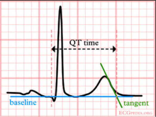
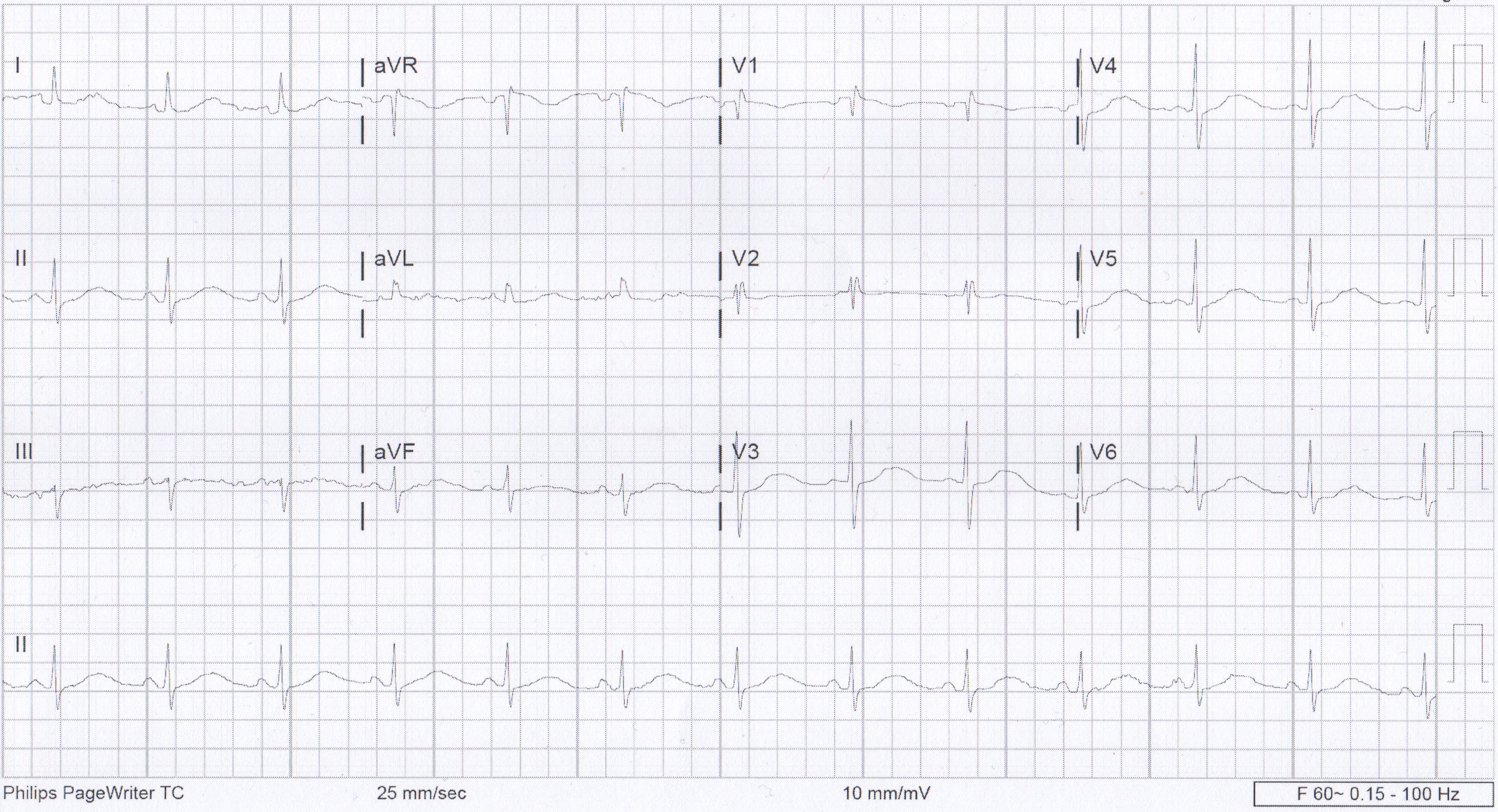
We usually talk about QTc rather than just the QT. This is because the QT interval varies depending on the HR. Using a correction equation standardizes the interval so it can be interpreted regardless of the HR. The EKG computer does give a calculation of the QTc, because we are obviously not hand calculating this on every patient. You should compare what the computer calculates to your gestalt when you review the EKG. If there are any concerns or discrepancies, you should hand calculate the QTc. MDCalc has an easy to use calculator. Above is the Bazett’s formula, which seems to be the most commonly used. Other formulas do exist. QTc is considered prolonged if > 440ms in men or > 460ms in women.

Next, why do we care?

ecg_hypokalaemia_torsades

This is the start of Polymorphic VT. There are several things that can cause this rhythm. Long QT is one possible cause. When Polymorphic VT is caused by a prolonged QT we give it a special name, torsades de pointes. The mechanism behind this is demonstrated on the above EKG. As the QT interval becomes more prolonged, there is a higher chance for an R-wave to hit on just the right part of the T-wave and cause this rhythm. QTc > 500ms seems be associated with higher risk.

So what causes prolonged QT?

Many things can cause prolonged QT. The most common etiologies are electrolyte abnormalities and drugs. Hypokalemia, hypomagnesemia, and hypocalcemia are well known to cause prolonged QT. Potassium and calcium are included on the CMP but don’t forget about magnesium. Drugs are also a big cause of prolonged QT. The list of drugs is long. Probably too long to memorize. However, there are some common medications and medication classes that you should know. The big classes are, Antiarrhythmics (like Amiodarone), Antihistimines (like Diphenhydramine), Macrolides (like Erythromycin), Antipsychotics (like Haloperidol), and TCAs (like Amitriptyline). This is not a complete list, just some highlights. If you really want to know if a specific medication is associated with prolonged QT, www.crediblemeds.org is a good source.

Other causes worth mentioning are structural heart disease, cardiac ischemia, and stroke. Some people do have Congenital Long QT Syndrome, but this should not be the leading diagnosis in the ED. Also those people are still at high risk for developing Polymorphic VT.

How did our patient do?

Her medications were reviewed and she was not on any of the most common offenders. Then routine labs came back. Unremarkable, except for K of 2.9! Repeat EKG after potassium repletion.

Normal EKG

EKG after K repletion


References:

  • www.uptodate.com
  • http://lifeinthefastlane.com/ecg-library/basics/qt_interval/
  • http://hqmeded-ecg.blogspot.com/2013/10/polymorphic-ventricular-tachycardia.html
  • bjsm.bmj.com/content/43/9/657/F3.large.jpgamp

Diabetes Insipidus in Intracranial Injury/Trauma

During a string of nights, I had two separate patients that despite having different injuries presented me with an interesting question. The first was a man in his 50s with a large intracranial hemorrhage, mass effect and the beginning of herniation; the second was a young male that was the victim of a GSW to the head. However, despite the different etiologies of their injuries, they both presented to Room 9 literally yelling for water. One became so combative in his demands for water, he had to be restrained.

This got me thinking; in the setting of very serious injuries, why is the only thing that concerns these men oral hydration?

The most concise information I found comes from Life in the Fast Lane. While it is referring to TBIs, the pathology relates to both of my patients as well. Thirst is controlled mostly by ADH released by the hypothalamus and transported to the posterior pituitary. Any disruption in this production chain can decrease ADH, leading to central diabetes insipidus.

In the setting of trauma, this can be caused by direct damage to the hypothalamus or posterior pituitary, disruption in their vascular supply or increased intracranial pressure/herniation that can compress these structures. Whether by a large hemorrhage or direct trauma like a GSW, intracranial injury can damage the ADH supply, leading to diabetes insipidus and the extreme thirst felt by these patients. Endocrinopathies have been associated with 30-50% of TBIs with the most common disorder being diabetes insipidus.


Just another overdose…..right?

20 yo M with unknown PMH comes in to room 9 with AMS and tachycardia to the 180s. Per EMS, he had been found down in his apartment, with crack cocaine pipes and other drug paraphernalia around him. He was found to be tachy as mentioned, as well as febrile with a temp of 103 axillary.

When he arrived in room 9, his HR was still in the 170s-180s. Blood pressure normal. He was pale, diaphoretic, and looked sick. He was speaking inappropriate words and would localize pain, GCS 12. Pupils dilated and briskly reactive. Rectal temp 104.1. CXR normal. Started IVF bolus and placed ice packs to the groin and axillae. Also gave Ativan as this was likely a stimulant overdose.

First EKG showed SVT at 180 BPM. After 2L of crystalloid and ativan, a repeat EKG showed sinus tachy at 140. The pt’s mental status was unchanged. The iStat showed a lactate of 13.

The plan was to place a rectal probe and monitor his temp, give him more fluids and Ativan prn, and re-assess later. I thought this was 100% an overdose. No problem.

About 2 hours and who knows how many room 9s later, I go to review his labs. I haven’t heard anything from nursing other than him continuously pulling out his rectal thermometer probe, so all must be well…Turns out he has a WBC of 44,000. Lactate has trended down, but he is still febrile to 102. This is when it hits me that maybe the guy who I’ve been treating for stimulant overdose is actually septic? His CXR and UA were normal, but maybe he has meningitis or encephalitis and that’s the reason for his mental status? Maybe I’m now 2 hours late with ABx?

I suppress the awful feeling in my stomach and go re-evaluate the pt. His mental status is unchanged from when I saw him in room 9. At least now his HR is in the low 100s. Given his mental status and tenuous vital signs, I know this patient is going to have to come in to the MICU. He’s going to need a head CT and an LP to rule out meningitis. I gave him antibiotics and called MICU. They evaluated the patient, and they agreed.

I chart checked the patient the next day. His LP was normal. His mental status improved overnight and he was transferred to the floor. Turns out this actually was likely all tox-related, but I thought it was a good learning point nonetheless. Sometimes it’s convenient to go down the path you’re led to by EMS or by nursing. Not only is it easy, but it’s usually the right path anyway. The stroke buzzer goes off and you immediately get your quick assessment over with so the patient can go to CT and stroke can do their thing. EMS tells you they found the patient in a house with drug paraphernalia, so you run with that.

But it’s important to keep the differential wide open when you first see a patient. At least consider less likely and less obvious possibilities. At some point, you’ll catch something that you otherwise would have missed until it was too late.