Antibiotic review

Maybe it’s because an overhead projector slide scanned and inserted into a powerpoint presentation comprised my medical school antibiotics curriculum (you know what I’m talking about if you went to U of L), but I’ve never really felt comfortable with the nuances of antibiotics. For those who want to understand them a little better, here’s a great review.

The bar is set, Chrissy!

NEXUS in the Elderly

Hopefully everyone is using the NEXUS criteria or the Canadian C Spine rule in evaluation of patients who have undergone neck trauma. Those familiar with both know one major difference, age criteria. NEXUS does not use age, Canadian C spine does. Using both rules together, like PERC with Well’s, increases sensitivity at expense of specificity.

Well here is a study on falls in the elderly (i.e. low mechanism which is another difference between NEXUS and Canadian) and application of NEXUS. Turns out, probably shouldn’t be using NEXUS in patients over 65. Liberally scan these folks, radiation is less of a concern, and the cost is justified due to morbidity of missed injuries. And of course do not bother with plain films (in adults).

Hot off the Press, Droperidol is Still Safe

Yet another quality article illustrating the safety of the most magical drug, droperidol. Not sure where people are getting it, as it is not being manufactured currently. We have none at any of my hospitals in Louisville. It is sad but perhaps someday we will get it back. In the same Annals issue an indictment of professional societies, journal editorial boards, and government advisory committees with their misinterpretation of “so-called facts.” Great reading, Dr Newman is the man.

Interesting case from the weekend – thoughts?

Hey guys, I was hoping to get your input on an interesting case I had at Kosair over the weekend.

16 yo F (6 ft, 150 lb…so basically an adult) with a PMH of depression, self injury, and prior suicide attempt presents after ingesting citalopram 40 mg x 90 pills (her prescription, just filled 2 days ago) and concerta 10 mg x 8-9 pills (her brother’s). Patient had been at a party the night before, admitted to EtOH.  Parents found out about the party the morning of admission and they had a big fight, took away car keys, etc. Patient decides to retaliate by swallowing pills, doesn’t tell anyone. Parents find her altered about 10:30, at Kosair at 11:40. Best guess is ingestion occurred sometime around 9-9:30am.  Had one seizure at home per family, and one en route per EMS. Generalized, tonic-clonic, brief.

Initial exam shows a drowsy but arousable patient. Answers orientation questions x3. Initial vitals show HR 147, BP 135/70, RR 25, 93% on some oxygen (can’t remember if NC or nonrebreather). Patient denies CP, palpitations, SOB, abd pain, N/V, weakness/numbness.  4mm, PERRL. MAE equally. Old self injury scars noted on wrists bilaterally. Exam otherwise unremarkable.

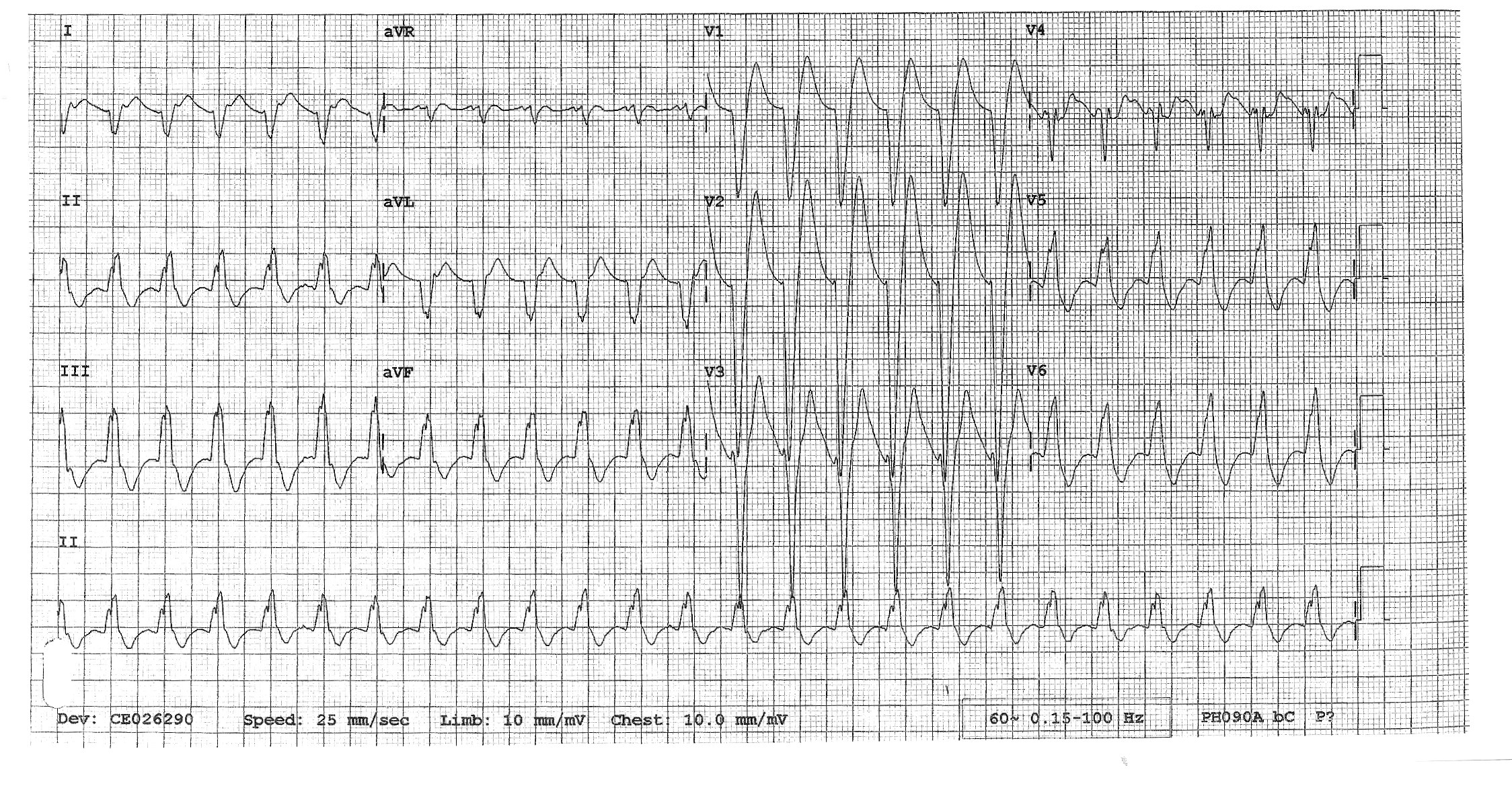
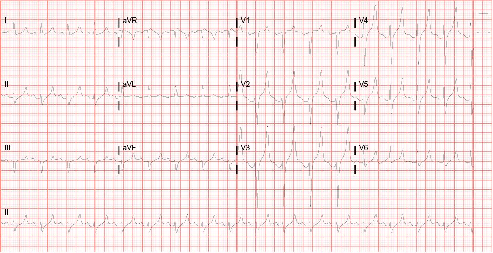
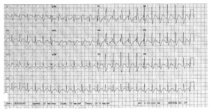
We start IVs, get her on a non-rebreather, get IV fluids going. Agree that charcoal seems like a bad idea with her mental status and seizures. Mom has shown up, and as we’re getting some additional history from her, respiratory is placing EKG leads. I’ve talked to poison control. Then, about 20 minutes into her stay, she seizes again. We bag her through the seizure, again generalized tonic-clonic, and just as we’re pushing 2 mg of IV Ativan she comes out of it. She appears post-ictal, but is maintaining her airway. We load her with Keppra, and as I glance at the monitor behind the attending’s head, I notice that her rhythm has changed and she looks like she’s got a wide QRS. We confirm she still has good pulses, still out of it mentally, and since she’s already connected to the EKG leads we grab one (time stamp 12:04):

EKG 1

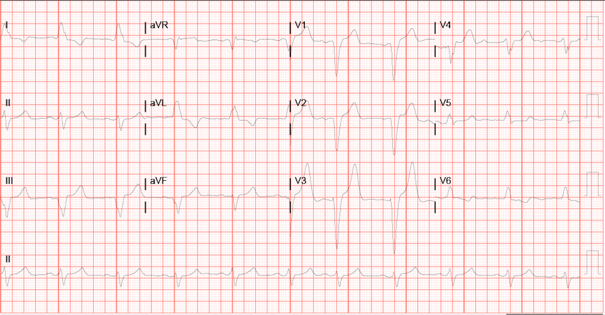
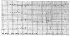
By the time we get this printed off (!!!!) she appears to have spontaneously converted back to sinus on the monitor. But woah, holy wide QRS/long QT batman! As the attending and I are pouring over the first EKG we get another one immediately (time stamp 12:08):

EKG 2

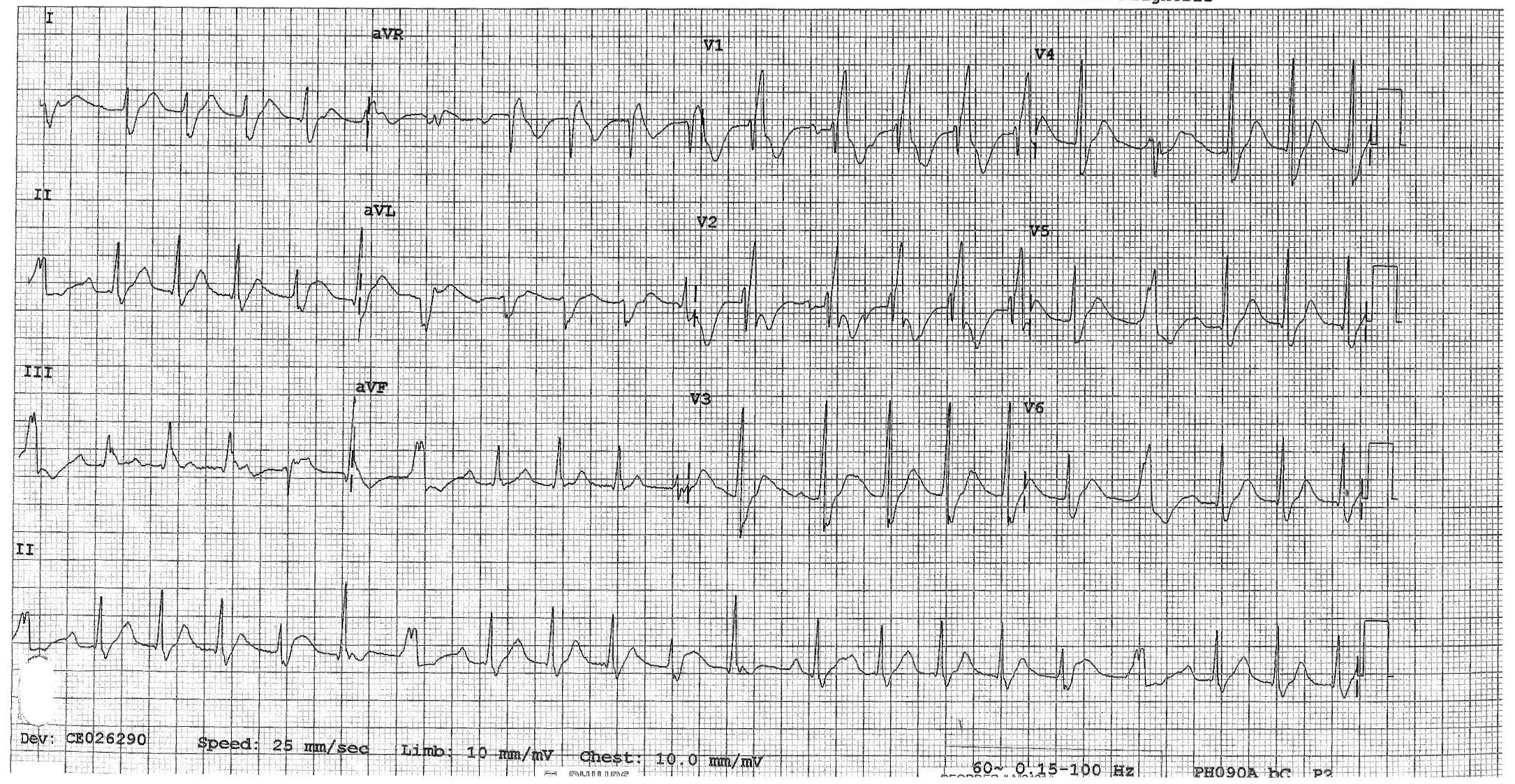
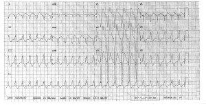
Thankfully, the QRS appears to have normalized, but we’ve still got a loooong QT, one of the things poison control definitely told us to look out for. Having seen a few similar ingestions at University, I suggest it’s time for bicarb. The attending wants to confirm and we quickly call poison control back, they agree and suggest starting a bicarb gtt, with pH goal of 7.45-7.55.  Now we look back at the monitor and she’s throwing a ton of PVCs, captured here on EKG #3 (time stamp 12:12):

EKG 3

At this point, we opt to push an amp of bicarb while we’re waiting for pharmacy to tube up the bicarb gtt. I have to say, we see it start to work pretty darn quickly. The PVCs slow down, and her rate really starts to head back towards normal. We get an iStat (shot me down when I suggested one earlier), and a few minutes after we’ve pushed the bicarb we get an initial pH of 7.15, pCO2 of 51.5, HCO3 of 18, BE -11, and AG of 21. Electrolytes were WNL. By this time we also know her pregnancy is negative, and her serum tox is negative, no acetaminophen/salicylates on board.  At this point, we talk about intubation as the patient’s mental status is still waxing/waning and she’s breathing shallowly with brief periods of apnea, almost like an opiate overdose. Attending wants to hold off, so I go off to call the PICU resident…and end up having to hang up the conversation halfway through when he changes his mind.

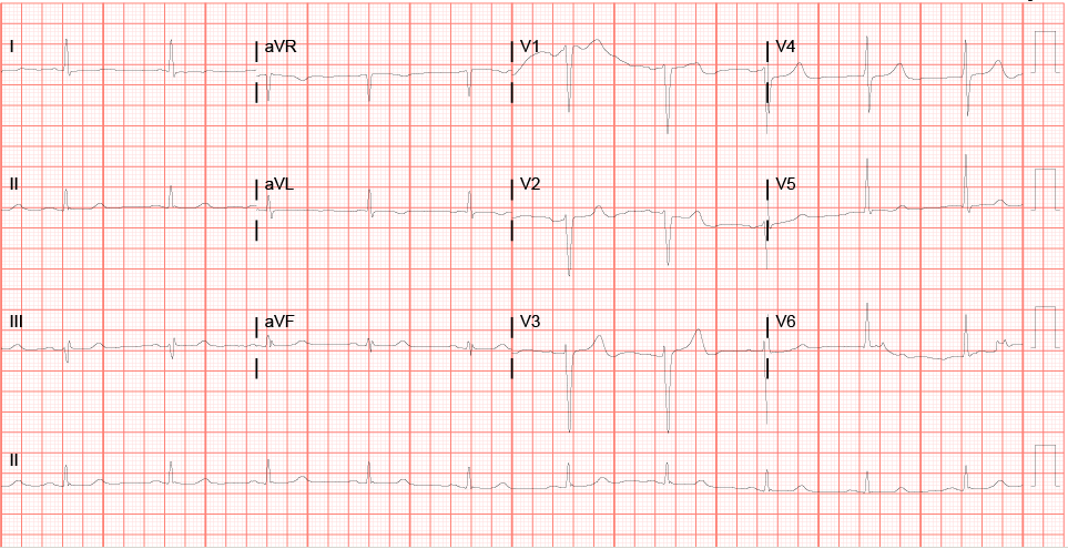
So we intubate her (finally got a peds tube…in an adult), the bicarb gtt comes up from pharmacy, they’re cleaning the PICU bed her, the last EKG looks 1000x better, and all’s well that ends well (time stamp 13:08):

EKG 6

So I’m curious to see what your all’s suggestions/thoughts are on this case.  Looking back at that first EKG, how would you classify it? We’ve got a wide complex, monomorphic tachycardia that to me looks like sustained V tach (with a pulse).  The long QT doesn’t surprise me, but this rhythm does as you’d typically you’d worry about it devolving into Torsades, but that’s not what this is.

Looking back, things I would have done differently:  get a temperature sooner/order a total CK (serotonin syndrome could have been a factor and we don’t have a recorded temp until she’d almost 2 hours into her stay, no one ever ordered a CK), intubate sooner, loading her with keppra when she hit the door after 2 witnessed seizures, maybe could have prevented the 3rd?

Also, if you’re curious, I found this “Toxicology Conundrum” on LITFL that specifically discusses citalopram overdose. Has some good info, citalopram is definitely one of the more potent SSRIs, and QT prolongation is dose dependent and can be seen after ingesting >600 mg (this chick took 3.6 GRAMS). Seizures are also fairly rare, only seen in 2-3% of cases.

Intractable Pain

I’ve seen about 3 cases now that presented with intractable pain. One was leg pain, put him in Room 9 much to the dismay of nursing, dude ended up having an acute arterial clot and received an amputation. One was acute on chronic back pain (history of chronic norco use, no change in quality) that ended up with an infected ulcer on his aorta (died later). And now this case….

67 y/o M presents with hip pain. Reports he was riding on a mower, hit a bump, had mild pain afterwards in his hip, but overall did well. He ambulated inside, took a shower, and after he sat down to do his business on the toilet he started to worsen. Especially when he stood up. No numbness/tingling/weakness/nausea/vomiting/fevers/chills/bowel or bladder incontinence. VSS. He has pin point tenderness behind his R hip. Worse with palpation. NO pain with axial load, internal/external rotation of leg. Pain with movement of torso. Pain isn’t nearly as bad when he’s just laying there. Appears in pain though at all times. Like TOO much pain.

Skip the XR knowing it won’t satisfy me, go straight to CT pelvis w/o contrast. It is normal. Decide to check labs at this point because I forsee an admission. He has a WBC count of 12. Cr of 1.8 (which is old) I admit him to the hospital for pain control as the idea of ambulation wasn’t happening, let alone just sitting up in the bed.

He is discharged the next day after MRIs of the L spine and hip are showing degenerative changes and lumbar stenosis and he is walking now (with the assistance of PT)

He presents again today and sees one of my partners. Again appears in pain, uncomfortable. Now has abdominal pain in the LLQ as well CT shows DIFFUSE pneumoperitoneum. He’s in the operating room at this moment…..

Sorting out theatrics/drama from reality can be a fine line we walk. Opiate abuse complicates things, and we often meet plenty of actors. But keep in mind pain out or proportion is almost always bad and delineating that can be difficult!

Always have your radar up, don’t be afraid to work up patients (regardless of what your ancillary staff and colleagues say)….. and don’t be afraid to be patient advocates and put them in the hospital to allow things to develop…..Remember One EKG, one CT, one lab draw, they are all stand still pictures of one moment in time and don’t always tell the whole truth. Certainly didn’t here in this case…..

Chest Pain Admission Dilemma

Hey guys here is an interesting article with actual patient oriented outcomes related to admission for chest pain.

Several take home points:

1. From highest quintile of admission rate to lowest (81% to 38%) the rate of MI and death went up by 3.6 per 1000 and 2.8 per thousand. This correlation implies that when you admit more patients you save lives.

2. It is VERY IMPORTANT to note the patient population. These are Medicare patients with average age of 71 years. So we ARE NOT talking about low risk chest pain ED patients.

3. Even though it looks impressive to save these lives, the NNT or number needed to admit to prevent one MI is 250 and to prevent one death is 333. Thats a lot of admissions. And admitting geriatric patients is often not a good thing for them. May be why the decrease in deaths is less than the decrease in MIs. They were dying because of a hospital acquired infection or deconditioning or something else.

4. It is striking to see how different the practice patterns are at different hospitals regarding admission of a fairly homogenous cohort of patients.

Appreciate any further comments.

EKG Changes in Hyperkalemia

I have had a couple of good EKG’s in the setting of hyperkalemia that I thought I would share.

The first case is a 68 y/o female with ESRD on dialysis presenting with “back pain”.  Turns out that her back pain was actually chronic and at baseline, but she had missed her last two dialysis appointments.  She denied any chest pain or SOA.  I ended up getting an EKG while waiting on labs.

Initial EKG:

Screen Shot 2015-08-04 at 2.46.53 PM

Previous EKG (~6 months ago)

Feb


What says you?  New onset LBBB?  After seeing this we gave Calcium Gluconate and asked one of our wonderful techs to grab a quick iStat as labs were taking forever to result.  Potassium was 6.8.  Gave insulin + D50 and bicarb.  EKG was repeated after Hyperkalemia treatment:

post

Renal consulted for emergent dialysis and medicine admitted.


Here’s another EKG that I had recently from a DKA patient who had an initial potassium of 7.8:

Hyper-K


Here’s a couple of good hyperkalemia resources:

Hyperkalemia EKG Basics

Treatment of Hyperkalemia

Just get a walking O2 sat

In patients with some suspicion for PE, even with a negative d dimer, I have often ordered a walking O2 sat and HR. This was not really evidence based, but maybe now could be. Below is the abstract for a prospective cohort study of patients known to be with and without PE. Interesting data even if only 114 patients. Cannot get full text yet.

Take home point. Combined sensitivity of HR increase of 10 BPM AND Sat decreased of >/= 2% was 100%.

ie if HR does not go up by 10 or more AND sat does not drop by 2 or more they are very unlikely, based on this small study, to have a PE.


CJEM. 2015 May;17(3):270-8. doi: 10.1017/cem.2014.45.

Ambulatory vital signs in the workup of pulmonary embolism using a standardized 3-minute walk test.

Abstract

OBJECTIVE:

Diagnosing pulmonary embolism can be difficult given its highly variable clinical presentation. Our objective was to determine whether a decrease in oxygen saturation or an increase in heart rate while ambulating could be used as an objective tool in the diagnosis of pulmonaryembolism.

METHODS:

This was a two-site tertiary-care-centre prospective cohort study that enrolled adult emergency department or thrombosis clinic patients with suspected or newly confirmed pulmonary embolism. Patients were asked to participate in a standardized 3-minute walk test, which assessedambulatory heart rate and ambulatory oxygen saturation. The primary outcome was pulmonary embolism.

RESULTS:

We enrolled 114 patients, including 30 with pulmonary embolism (26.3%). A ≥2% absolute decrease in ambulatory oxygen saturation and an ambulatory change in heart rate >10 beats per minute (BPM) were significantly associated with pulmonary embolism. An ambulatory heart rate change of >10 BPM had a sensitivity of 96.6% (95% confidence interval [CI] 83.3 to 99.4) and a specificity of 31.0% (95% CI 22.1 to 45.0) forpulmonary embolism. A ≥2% absolute decrease ambulatory oxygen saturation had a sensitivity of 80.2% (95% CI 62.7 to 90.5) and a specificity of 39.3% (95% CI 29.5 to 50.0) for pulmonary embolism. The combination of both variables yielded a sensitivity of 100.0% (95% CI 87.0 to 100.0) and a specificity of 11.0% (95% CI 6.6 to 21.0).

CONCLUSION:

In summary, our study found that an ambulatory heart rate change of >10 BPM or a ≥2% absolute decrease in ambulatory oxygen saturation from baseline during a standardized 3-minute walk test are highly correlated with pulmonary embolism. Although the findings appear promising, neither of these variables can currently be recommended as a screening tool for pulmonary embolism until larger prospective studies examine their performance either alone or with pre-existing rules.

Dr. Smock’s Forensic GSW lecture 7/22/15

Here are some highlights of Dr. Smock’s Forensic lecture. This will help remind me what to document in my next GSW pt. Here is a pfd version. Forensic GSW Documentation


Forensic GSW Documentation: 

Bullet causes… Abrasion collar
Unburned gunpowder causes… Tattooing aka. stippling (this lasts a few days, seen as punctate abrasions, DO NOT CALL THIS “GUN POWDER”)
Burned gunpowder causes… Soot
Flame causes… Seared skin
Injected gas causes… Triangular tears
Muzzle causes… Muzzle contusion

Distance from Weapon:

Indeterminate abrasion collar present
Intermediate < 40 in Tattooing & abrasion collar present
Close < 6 in Soot & abrasion collar present
Contact Seared skin, triangular shaped tears, & soot present

Straight to the cuff

To get us and RT on the the same page with stylet shaping in ETI.

First, what does our text book say about it:

Screen Shot 2015-07-28 at 8.10.24 PM Screen Shot 2015-07-28 at 8.11.19 PM

In case anyone doesn’t recognize this, it’s from Robert’s and Hedge’s Procedures in Emergency Medicine. This is the “other” textbook, that you absolutely have to read.

For some more on this topic:

http://www.epmonthly.com/features/current-features/avoiding-common-laryngoscopy-errors-part-ii/

This is from Dr Rich Levitan. He is the king of the airway, and I highly recommend his book, The AirwayCam Guide to Intubation.

For the interested learner, or those to lazy to read the above, here are some video examples:

Finally, if anyone feels the need to brush up on intubation in general, here are two more of my favorite resources.

Scott Weingart

And the omnipresent Life in the Fastlane:

http://lifeinthefastlane.com/own-the-airway/

JG MD


Snakebite

Recently saw at case at ULH (Not mine but posted with permission from resident involved) of a young female (18-20yo) flown from OSH for copperhead bite to L foot. Pt had been hiking in the woods with her boyfriend when she felt a stabbing sensation on her foot, looked down and there was a snake. Pt took a picture of it – sure enough it was a copperhead. Labs at OSH wnl, sent to ULH for concern for possible need for antivenin. I realized – being an urban trauma center – we don’t see a lot of snake bites. If you’re from the area, you may not be familiar with our venomous critters.
There are four species of venomous snakes in KY, all pit vipers (triangular heads, heavy bodies, cat-like pupils) – fw.ky.gov
1. Copperhead – extremely common throughout the state, most common envenomation, mildest venom of the four. (As an acquaintance of mine, Jim Harrison of East KY Venom Extraction lab used to say – no recorded history of death from copperhead envenomation in KY history
2. Timber Rattlesnake – found throughout state, potent venom, relatively docile. Dark coloration, have a rattle (obviously)
3. Pygmy Rattlesnake – found in extreme Western KY. Also have a rattle, potent venom
4. Cottonmouth – found in Western KY, potent venom, can be aggressive. Can be mistaken for Eastern Water Snake (common, non-venomous, aggressive water-dwelling snake found throughout KY)
Best identification – hopefully someone took a picture – otherwise, one to two small puncture wounds with increasing swelling and pain are good signs of envenomation. However, approximately 25% of viper envenomations can be dry bites – where snake gives a warning bite, injects no venom.

Signs and Symptoms of snake bite:
Venomous snakes in KY typically have venoms containing cytotoxins and hemotoxins – they break down tissue and can act on coagulations factors
-puncture wounds (usually paired, can be one)
-erythema and swelling
-intense pain at site and increasing proximally
-systemic symptoms can include nausea, vomiting, abdominal pain, vertigo. Altered mental status and hypotension can present as well

First Aid if bitten:
-Walk slowly – increased activity increases circulation of venom
-Elevate affected part: most bites are on distal extremities: feet, hands. More serious complications are associated with bites involving trunk, neck, face
-Wash with soap and water
DO NOT:
-Apply a tourniquet
-Attempt to suck venom from wound
-Apply ice to wound

Medical Work-up for known or suspected envenomation
-ABCs (always the first step)
-Good H&P – observe amount of swelling, movement of affected area. Mark swelling and erythema with marker to observe for progression
-CBC
-Comp
-Coags
-Total CK
-Fibrinogen
-U/A – to look for myoglobinuria
-Consider ABG and lactic acid if signs of systemic toxicity
-Xray to r/o retained teeth

Severe complications
-Rhabdomyolysis – from muscle breakdown from venom
-Compartment syndrome – cannot be diagnosed on clinical exam alone. Venom can cause tingling and paresthesias. If concerned, consult your surgeon
-Thrombocytopenia and coagulopathy – caused by thrombin-like molecules in venom. Treated with antivenom
-Systemic toxicity – can be life-threatening

Treatment:
-Update Tdap if indicated
-Pain control, elevation
-Antivenom if indicated

Antivenin: Always contact your toxicologist prior to giving
-Recommended for moderate to severe envenomations
(http://www.uptodate.com.echo.louisville.edu/contents/image?imageKey=EM%2F53948&topicKey=EM%2F6595&rank=2%7E39&source=see_link&search=snake+bite&utdPopup=true)
-Dosing: Moderate envenomation – 4 to 6 vials (1g each) over 60 min with repeat dose as needed
Severe envenomation – 6 to 9 vials over 60min with repeat dose prn
-Contraindications: papaya allergy (contains enzyme papain)
-Stop if signs of anaphylaxis

Disposition
-Requires antivenom – admission. Complications can include compartment syndrome, delayed coagulopathy (up to 48hrs). May need redosing of antivenom
-Mild envenomation or no symptoms – can be discharged home if pain controlled and no signs of toxicity at 8-12hrs s/p bite. If bitten by rattlesnake or cottonmouth, recommend CBC, PT, fibrinogen at 2-3d and 5-6d after initial bite