Ready for discharge?

Interesting case from my MICU month.  Pt is a middle aged  WM with hx of HIV, CVA presented to the ED with hypoxic respiratory failure and sepsis. Intubated in the ED and admitted to the unit. Pt self extubated few hours later in the MICU. Responded well to fluids, antibiotics and O2 nasal cannula.  Few days later, nurse d/c’ed his RIJ central line.

Within minutes,  pt became severely altered, O2 sats plummeted to the 60s and  bedside ultrasound showed significant air bubbles in the cardiac chambers.

What happened? When central line was removed, the wound site was not immediately occluded (with fingers, dressing with tape, etc). As pt inhaled, air traveled through the communicating conduit into the central blood vessel.

What to do now? Intubate for severe respiratory distress. Place pt in left lateral decubitus position and Trendelenburg position–prevents air from traveling to the pulmonary arteries causing airflow obstruction. Hyperbaric oxygen therapy, if you suspect cerebral embolism, as it decreases mortality.

The pt was intubated, placed on his side with bed tilted down. Did fine and transferred to floor the next day.

Positioning is Everything

When using a chest x-ray to look for a pneumothorax, positioning of the patient is everything.  The first chest x-ray below is an upright chest x-ray from an OSH of a patient that fell 30 feet from deer stand and was found to have a right pneumothorax.  The OSH didn’t do any other imaging and didn’t even send the patient with a c-collar.

When the patient arrived we laid him down and placed a c-collar and assumed that his spines weren’t cleared yet.  When we shot the portable, supine chest x-ray in our ED we couldn’t see a pneumothorax and our radiologist actually read it as no pneumothorax.

Using the US, an EFAST was performed and showed a pneumothorax and the subsequent Chest CT verified it.  Therefore the next time you get an ED, supine chest xray on someone, remember that just because you don’t see a pneumothorax on a supine CXR, doesn’t mean they don’t have one.  The optimal xray is an upright chest xray (expiratory if possible)!

Upright OSH xrayUpright Chest x-ray from OSH

Supine UofL portable Xray

Portable, supine Chest X-ray in our ED

CT scan

CT showing the Right Pneumothorax

Acute Cholecystitis, Classic

Interesting case from a couple weeks ago.

20ish yo white male, no significant past medical diagnoses. Overweight. Family history of gallbladder disease. No OTC or Rx medications. Patient smokes, does not drink, and has used IV drugs in the past.

Here in the ER due to RUQ abdominal pain for one week, was coming and going and is now constant. On further questioning, admits that his mom made him come because of her history of gallstones and cholecystectomy. On exam patient has jaundiced sclera and urine on table is dark brown, pain in right upper quadrant of abdomen is exquisite. States he has been vomiting especially after eating and all food makes him sick. No documented fevers, but feeling chills.

Urine: Large bilirubin, otherwise normal

Pertinent blood: WBC 8.4, Hgb 14.2, Plt 430. Alk Phos 140, AST 734, ALT 1417, Total Bili 9.5. Lipase 22. Tylenol neg.

Didn’t expect this guys ‘acute chole’ to cause liver failure.  Either way, he was getting further imaging to find out more. No ultrasound coverage at 5am so CT for now, then ultrasound at 7am. Added tox screens and hepatitis panel at this time.

CT abdomen/pelvis with contrast: “Markedly thickened/edematous wall of the gallbladder indicating cholecystitis. No calcified stone visualized. Additional imaging maybe obtained with ultrasound.” Also, normal liver.

The results of the ultrasound showed a “nondistended gallbladder with marked wall thickening, edema and a positive sonographic Murphy sign. Given lack of clear visualization of the posterior wall, highly worrisome for complicated cholecystitis, possibly gangrenous or with a focal posterior perforation.” Normal liver and mildly dilated bile duct.

Now with labs showing liver failure and two forms of imaging showing acute cholecystitis, it had to be. Admitted to the general surgery team though the ‘acalculous cholecystitis’ with liver failure was enough to peak my interest in follow-up.

Hepatitis panel comes back later same day showing reactivity for Hep C. Discharge 5 days later, no surgery, no acute interventions, with down-trending liver function panel and follow-up with the GI clinic.

While most commonly associated with cholecystitis, a quick literature search reveals multiple reasons besides cholecystitis to have gallbladder wall thickening… congestive heart failure (right sided), gallbladder carcinoma, adenomyomatosis (chronic gallbladder inflammation or degeneration), renal failure, pancreatitis, cirrhosis and other forms of liver failure.

Just some thoughts on stroke management in the acute setting…

A couple months ago (during lecture), we had a discussion regarding tPA and acute onset of stroke. As you would expect, we discussed the indications, contraindications, etc of treating stroke with tPA. We also touched on the subject of our role in pushing tPA here at UofL.

Obviously as a stroke center, our stroke team is working around the clock- and as such, generally takes the ball on this one. However, when we are practicing outside of UofL, a stroke team is not always going to be available and ultimately the management will fall on us. Hence the discussion and considerations for us making the call or at least working towards that possibility in the future.

Coincidentally, at precisely the same time as this conversation, a small journal, based out of New England, with a primary focus on medicine, published three fine articles on a similar topic. These articles were focused on the treatment of stroke in  the acute setting.

As pertinent to this post, they explored the use of thrombolysis as well as mechanical removal of thrombus when said thrombus is located in a proximal vessel. Now I won’t pretend I can read, but for those of you who can- below, are links to two “previews” and one complete view of the above mentioned articles. Additionally, an audio file is embedded, containing a break down of the articles by Dr. Dave Newman, of the Mt. Sinai School of Medicine in the Department of Emergency Medicine, to be featured on a future episode of EM:Rap.

Ultimately, I guess the questions I have regarding this as a post on Room9er are as follows:

1. I believe the Stroke team has embraced these articles and to my knowledge (as of February) may have moved towards mechanical retrieval of thrombus in proximal vessels in appropriate candidates. If we are trying to move towards a more ED involved decision tree, what will we need to know and where will our policies stem from? At least as a concept. At this time, I have only heard us discuss tPA, but if we as a hospital are moving towards multiple modalities for treatment of acute stroke, should we not be discussing these as well?

2. With consideration of a 6 hour time frame to thrombus retrieval, what is our (UofL) policy on timelines regarding retrieval. (This is not reflecting any current policies, merely one parameter from one study.)

3. How much time is required from page to cath? This is undoubtedly a big question. What does it take to have a vascular team, NES team, etc ready do go. How will this influence our time of onset to treatment guidelines.

4. Outside of UofL, taking into consideration transit time, etc. how will this influence the management of stroke? Based on CTA availability, transit time, local resources, etc.

ESCAPE

EXTEND_IA

MR CLEAN


Changes to tPA Contraindications in Acute Ischemic Stroke

Since its approval in 1987, controversy has surrounded a drug that we all know is near and dear to my heart, recombinant tissue Plasminogen Activator (insert eye roll).  In similar discreetness of a Hollywood wedding, the FDA updated the prescribing information of tPA with important changes made to the contraindications to the use of tPA in the setting of acute ischemic stroke.  It is unclear as to what prompted these updates and why.  There have been no recent studies of significance published to support these modifications.


2/2015 updated prescribing info:

“Do not administer Activase to treat acute ischemic stroke in the following situations in which the risk of bleeding is greater than the potential benefit:

• Current intracranial hemorrhage

• Subarachnoid hemorrhage

• Active internal bleeding

• Recent (within 3 months) intracranial or intraspinal surgery or serious head trauma

• Presence of intracranial conditions that may increase the risk of bleeding (e.g., some neoplasms, arteriovenous malformations, or aneurysms)

• Bleeding diathesis

• Current severe uncontrolled hypertension”


Now contrast the new package insert to the 2013 package insert.  Pay special attention to the omissions of the exclusion of contraindications in patients with history of intracranial hemorrhage and seizure at onset of stroke.   It is pertinent to note that the wording regarding the contraindications has also changed.  The previous consequences being “significant disability or death” have now been replaced with “situations in which the risk of bleeding is greater than the potential benefit.”


From the 2013 package insert (changes are italicized):

“Activase therapy in patients with acute ischemic stroke is contraindicated in the following situations because of an increased risk of bleeding, which could result in significant disability or death:

  • Evidence of intracranial hemorrhage on pretreatment evaluation
  • Suspicion of subarachnoid hemorrhage on pretreatment evaluation
  • Recent (within 3 months) intracranial or intraspinal surgery, serious head trauma, or previous stroke
  • History of intracranial hemorrhage
  • Uncontrolled hypertension at time of treatment (e.g., > 185 mm Hg systolic or > 110 mm Hg diastolic)
  • Seizure at the onset of stroke
  • Active internal bleeding
  • Intracranial neoplasm, arteriovenous malformation, or aneurysm
  • Known bleeding diathesis including but not limited to:

o       Current use of oral anticoagulants (e.g., warfarin sodium) or an International Normalized Ratio (INR) > 1.7 or a prothrombin time (PT) > 15 seconds

o       Administration of heparin within 48 hours preceding the onset of stroke and have an elevated activated partial thromboplastin time (aPTT) at presentation.

o       Platelet count < 100,000/mm3”


The new 2/2015 update does provide some vague conditions in which the risks of bleeding must be outweighed against the anticipated benefits (however, note that these are not firm contraindications):

• Recent major surgery or procedure, (e.g., coronary artery bypass graft, obstetrical delivery, organ biopsy, previous puncture of noncompressible vessels)

• Cerebrovascular disease

• Recent intracranial hemorrhage

• Recent gastrointestinal or genitourinary bleeding

• Recent trauma

• Hypertension: systolic BP above 175 mm Hg or diastolic BP above 110 mm Hg

• High likelihood of left heart thrombus, e.g., mitral stenosis with atrial fibrillation

• Acute pericarditis

• Subacute bacterial endocarditis

• Hemostatic defects including those secondary to severe hepatic or renal disease

• Significant hepatic dysfunction

• Pregnancy

• Diabetic hemorrhagic retinopathy, or other hemorrhagic ophthalmic conditions

• Septic thrombophlebitis or occluded AV cannula at seriously infected site

• Advanced age [see Use in Specific Populations (8.5)]

• Patients currently receiving anticoagulants (e.g., warfarin sodium)

• Any other condition in which bleeding constitutes a significant hazard or would be particularly difficult to manage because of its location.


Currently the guidelines have not updated their list of contraindications to tPA in acute stroke, but I wouldn’t be surprised to see them included when the guidelines are updated.


So like Oprah says, you get tPA! You get tPA! Everyone gets tPA!

Sinus tach Part II

This is a case that ended up being a 72 hour return. Another good example that sinus tach should have a good explanation. Another good reason to know discharge vitals!

Middle aged M, history of TBI, PE, MRSA bacteremia, s/p trach/g-tube/IVC filter, presents with displaced g-tube and increased agitation. Trauma consulted, g-tube replaced. Fluro shows good placement. Exam otherwise unremarkable. No labs performed. Documentation of to be non-verbal/not following commands and this was baseline. VS prior to discharge. Normal BP, HR trend: 68->70->115->111 (at discharge)

Patient returns for changed mental status. Not documented in what way he was changed from baseline. But found to have BUN: 115., Cr: 2.96. BP 90’s systolic, HR wnl. Head CT negative. Hgb: 12, INR: 1.6 (on coumadin) Na: 154, Cl: 112. Otherwise labwork unremarkable.

Patient admitted to medicine service. Hgb trended down 12.4->9.8->8.4. Patient receiving IV fluids during this time. Mild improvements in BUN/Cr. Patient was a STAT response 5 days later. Hypotensive/tachy. Hgb: 5.3 Dark stools noted. Transferred to MICU. GI scoped, found to have erosion of IVC filter into duodenum. Vascular consulted. Patient transfused/stabilized. IVC filter removed, transferred to floor. BUN/Cr normalized during stay.

Two things here, the HR as mentioned, should always have a good explanation. In a patient like this, the history is limited, more information is probably useful than less. Granted a patient like this is very difficult to evaluate at baseline, I’d lean towards shotgun labs/imaging etc. Not sure if it would’ve made a difference in the end but nonetheless.

On the 2nd visit though, a BUN of 115 should raise an eyebrow That’s a BUN/Cr ratio greater than 20. This patient could be just a simple AKI due to hypovolumia. But a BUN that high should also raise the suspicion of a GI bleed. A hemoccult probably is indicated at this point (for somebody that can’t relay much information at baseline). I don’t think anybody would’ve predicted the cause, but nonetheless, neither any of us, nor IM really interpreted that BUN as it should’ve. Just a few notes on Bun/Cr below taken from life in the fast lane. Not definitive, but just something to do a double take on.

Urea:Creatinine Ratio (in the setting of renal failure / elevated creatinine)

20:1 – normal or post renal cause of AKI
>20:1 – pre-renal cause (urea absorption increased compared to creatinine)
<20:1 – intrinsic renal damage (urea unable to be absorbed -> become like creatinine -> ratio gets closer to 1)

Sinus Tach Part 1

Abnormal vital signs are always something to keep an eye on. Always important to explain it in documentation, but keep an eye on the trend throughout your shift as it’s one piece of information that might tip you off that something else may going on. Try to resolve the vital signs prior to discharge, looks better from a chart buffing standpoint, but also an indication that you have corrected the original problem. And if you can’t, again explain it, or just don’t discharge the patient. You will never be 100% in this job, and your initial impression could be flat out wrong, so putting in little safeguards to protect yourself, from well yourself, can help you not miss things.

Quick case…..47 y/o M, history of 75% TBSA burns, active c-diff, PNA presents to the ED for low Hgb. Had Hgb of 7 at NH, 6.4 when checked here. Was just discharged recently from our ER (by myself, VSS at d/c) with new blisters on L scalp, Hgb was 7.3 at that time, 7-7.8 on prior visits. Wbc count: 11.8. Chronic indwelling foley. Urine dip: blood/wbcs/bacteria, neg leuk esterase/nitrite, already on levaquin for known PNA as well. Sinus tach to 120s. Received transfusion x2 units and initial plan was d/c back to nursing home per initial resident. Checked out to 2nd resident (myself) HR remained tachy to 120. Medicine consulted for admission. He was Afebrile. Blood cultures/urine cultures/ sputum cultured ordered as he did have an elevated WBC. Broad spectrum abx ordered. Rectal temp ordered x2, which were both normal. Medicine consulted for low hgb, sinus tach, and they cancelled cultures and abx, felt it was due to anemia although it did not improve with 2 unit transfusion (they are not always right). Planned for anemia work up and obs.

The patient admitted to the hospital and transfused. Hgb trended down again and patient required transfusion again on 9/26. Neg hemoccult/GI bleeding. Anemia work up not completed, but appeared to likely be of chronic disease vs iron deficiency. Patient became febrile on 9/23, BP dropped (that’s 3 days later…..) Started on broad spectrum abx, pan cultured. Urine grew acinetobacter. Blood cultures neg/c-diff negative. Hypotension was not responsive to fluids. Palliative consulted, patient and family elected to be comfort care. Made DNR. Comfort measures in place, was to be discharged to hospice. Patient subsequently expired prior to discharge on 10/4, etiology believed to be sepsis.

This patient had obviously a lot going on. Sinus tach could’ve been from anemia, pain, sepsis, stress, anxiety, etc. The initial thought it was due to anemia. but if this was true, it should come down with fluids and/or blood? Or at least partially respond? But it didn’t budge. He’s obviously had a long in-house history with the history of the burns so he was set up for a resistant bug, odd presentation. And honestly when we start seeing PNA + UTI we should start leaning towards sepsis from 1 agent with seeding, rather than a PNA and a UTI as two different infections going on. But in the end when he was checked out to me and the HR hadn’t budged, that was the tip off that something else was going on.

To expand on this as you can see things weren’t handled as they should’ve been initially. I see some people use the medicine service as a consultant. Which I thoroughly don’t believe in. They practice essentially the same thing as us, except less broad, more in depth, and a hell of a lot more boring (yes offense). When we talk to NES or neurology, or OB, or any of the other sub-specialties we are generally looking for guidance and information about a practice that is beyond our skills, beyond just basic medicine. And they have texts/papers/experience/OR time that allows them to function as a consultant and specialist in their area (especially the surgical services!).

But the medicine service is not all that different from what we do. When we call them, it shouldn’t be, “come see if this guy is ok for discharge.” We know which ones are and aren’t. And if you don’t, read more, use 5-minute EM consult, follow up on the patients you admit, so you can get a better understanding of what happens beyond the ED. You might not know the entire algorithm for hyponatremia and what work up they exactly do, but you should know when to admit one and how to emergently treat it. Or when to transition from insulin gtt to metformin, but you know when the glucose is too high to dispo home. Etc etc……. In their defense, sometimes we call them with patients that can theoretically be managed at home to begin with, but don’t fool yourself into thinking they know exactly what can go home and what can stay, and that they are the almighty, all-knowing doctor (case in point above.) They are residents too, and they don’t have an attending looking into them real time like you do (they have uptodate, which I can only assume why it takes 6 hours to admit asthma or whatever.)

Anyways to get back to my points.
1.Keep a close and scrutinizing eye on those VS.
2.And don’t let medicine pretend to be smarter than you.

Case #2

Case #2:

30 yo F h/o morbid obesity and DM2 not well controlled on insulin and metformin presents after high speed MVA vs pole. 5 month old baby in back carseat sent the the Ped’s ED, unharmed. EMS called, prolonged extrication about 30 min, vitals en route stable, BP 120/76 and HR 96 just PTA (hmm?). No IVs established, axox3, talking, calm and cooperative.

On exam, breath sounds normal, seat belt sign obvious on lower abdomen with mild LUQ abdominal tenderness on exam, main c/o left thigh pain. Appears twisted but unsure if broken. Patient is morbidly obese, probably 350-400 lbs, which causes some problems next: placed on the monitor and HR 130, check pulses and does have pulses in distal extremities though weak, BP unable to get multiple times, then manual BP unable to obtain. Ask nurse why no BP, states “cuff not big enough to work properly,” also trying on leg.

IV placed in right AC no problem, placed NS on pressure bag. HR during xrays comes down from 130–>115.

CXR done nothing obvious, PXR done, again nothing obvious and left femur show proximal 3rd shaft fx, traction splint placed by intern, FAST scan neg in cardiac window, but very positive in pelvic window as well and slightly positive in RUQ and LUQ. Level 1 called after FAST and finally a BP obtained 80/40 as trauma walks in the door!!! Patient no longer with palpable peripheral pulses, good central pulses, a&ox3.

Ever feel like an idiot…just watched this lady with HR 130 and BP 80/40 for 18 minutes prior to level 1 trauma call!

Blood, central line, another peripheral IV. Trauma dawdled a little in ED more than should have, repeated the very positive FAST scan, though we did resuscitate her with blood and fluids, central line, trauma attempted a-line (apparently they didn’t trust the very low BP either) and finally went to CT scanner (another 25 min) and thought to have SMV avulsion (yikes) prior to taking her to the OR.

Diagnosis: avulsion of superior mesenteric artery (even worse). left femur fracture.

Patient spent more than one month in the SICU, never extubated, multiple loops of bowel resected for necrosis and never closed her abdomen, family decided to withdraw care after she continued to go downhill and quality of life would have been an issue.

Let me point out the obvious, I should have called a level 1 earlier. 10 min earlier when  had HR in 130s and unable to obtain BP x 2 would have been enough. Unsure if her outcome would have been different, but sure makes me wonder.

Often times it is hard to get a BP on a morbidly obese person, esp when have peripheral pulses and axox3, but it is much better to be safe than sorry, call the level 1 when in doubt. Better for the patient’s sake to feel like an idiot earlier than to feel like an idiot later in the game.

The other thing is, we had her packaged for the OR when trauma arrived, peripheral IV and blood obtained, 1L fluids, HR 110, CXR, PXR and femur, traction splint to femur, FAST done and very positive. Should have gone straight to the OR as soon as trauma arrived. Trauma fellow wanted CT scan and further resuscitation (why try an a-line?) which took time.

Feedback and comments appreciated.

Couple of recent cases…

One of the most interesting posts recently was Zach’s post about what he could have done differently. Thought I would continue this trend and post a couple of cases that could have gone better and leave some things up for discussion. Case one here and two next post.

Case #1: 90 yo F h/o HTN and arthritis, restrained passenger of moderate speed MVA, driver was unharmed but car was going too fast and struck the back of a semi-truck. Extrication about 10 minutes. Patient c/o right leg pain and right forearm pain, skin tear to right arm and obvious Colles fracture and femur fracture likely as well on initial exam. No chest or abdominal pain. Kept in R9 for the potential femur fracture and age. Vital signs all been stable, patient a&ox3, talkative and despite mild pain appears in good spirits.

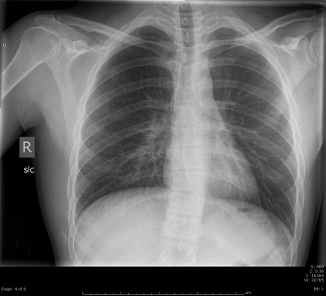
On initial exam, HR 96 and regular, 125/70, RR and temp normal. Pain controlled as long as not moving. Good breath sounds, minimal chest tenderness on palpation of sternum, abdomen and pelvis unremarkable. Placed in traction splint in R9 for significant pain and shortening of right leg. CXR normal, PXR normal, and R femur with proximal femur fx minimally displaced. FAST neg. Give tdap and bag of LR. Further eval shows pain in right leg from femur/knee/tib/ankle and also left femur/knee/tib. As mentioned obvious fx right Colles, with pain in right elbow/forearm/wrist/hand and  also pain left forearm and wrist. That makes for man scan + a whole ton of xrays in all extremities.

Spoke with ortho on the phone in R9 due to known femur fracture. Vitals on R9 exit unchanged. To the CT scanner. Nurse calls from xray after CT done and asks for small amount pain meds as patient now c/o more pain, especially in right leg and arm. Dilaudid 0.5mg ordered. Patient comes into main ER literally 90 minutes after initial presentation due to so many xrays! (This is my fault). Labs unremarkable, Hgb 11. I see the CT scans but nothing obvious to me, no head bleed, no c-spine fx, no PTX, no obvious free fluid in belly. See patient as she as she comes back bc ask for more pain meds. HR now 120s, irregular?, BP 95/65. Ask nurse how long this has been going on, states “oh, just the last 30 min or so.” Ortho at bedside as well, wants to do sedation.

EKG done, show afib rate 120s (no h/o afib). BP cont to be 90/60s. Giving 2nd L LR, nurse start 2nd IV and get L NS going. Patient still talking and states she feels ok other than the pain in her leg. Call to Trauma (should have called earlier in 90 yo with known femur fx), but by now its been more than 100 min total time in ER. Trauma arrives pretty quickly, patient now been here 2 hrs. Agree with fluid boluses, talk about patient condition with fellow and wedge and all agree think likely due to trauma in afib and that why BP sucks.

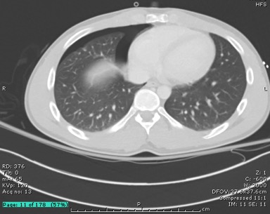
Tell ortho that too high risk patient and unstable to do sedation. Does hematoma block right Colles fracture with reduction as well and leaves the femur in traction. Admitted to SICU after just over 2 hours in ER. CT head and c-spine only scans back and are neg at admission. About 30 min later rest of man scan comes back. Small nondisplaced sternal fx and very small hematoma (but possibly active bleeding) to right retroperitoneal area. Again speak to trauma about this. Asked about the retroperitoneal bleed and fellow states it very small and should wall off without intervention based on place. Possibly afib due to contusion? Cards was consulted by Trauma who saw patient and said to control pain, resuscitation and other trauma factors (unhelpful but true, not much for them to do).

Brings up a question I had, in setting of trauma, what medicines should be given for afib with RVR? Cardiac contusion? Or just let it ride? Trauma asked the nurse to give metoprolol but this was never given mainly due to nursing concerns about BP.

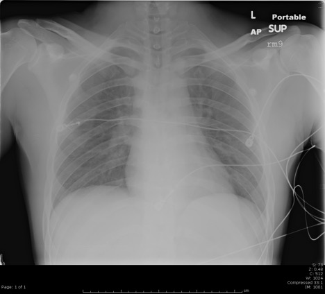
Ok, long story short, HR cont to be 120-130s, afib, BP 90/60s, after being ICU hold for about 2 hours patient had decreasing mentation. Trauma placed central line and a-line and ABG showed pH 6.8! Istat Hgb 8.0. Blood given, minimal response.

Patient intubated by trauma, arrested during intubation, one round CPR and came back, went to SICU same vitals, called in IR and intervention showed minimal bleeding, thought to likely be venous, but did have several coils placed. More blood and then pressors through the night. Arrested 2 more times throughout the next 6 hrs, family still wanted all interventions. Finally about 12 total hrs after ER presentation, arrested for about 20 min and TOD called.

Couple of main points from this case that I learned and hopefully helpful for you all:

1. As Coleman says, old people go down hill quickly, be on your guard no matter how good they may look on presentation.

2. Be careful of ordering too many xrays, me not seeing the patient for over an hour while in xray is unacceptable, I should have known the change in her vitals either by seeing it directly or have nursing tell me.

3. I knew this was a trauma admit as soon as she hit the door being 90 yo and femur fx, but I didn’t call them for a long time, why? Bc I didn’t have much to tell them (couple xrays, stable VS, man scan taking 3 hrs to get read). But just be sure to get them involved early.

4. We don’t typically repeat labs or get istat too often, but for someone like this who takes a turn for the worse, would have been worth it to get hgb after her vitals became unstable.

5. Her mentation decreased after admission, but I could have been more aggressive with central line and resuscitation (blood), she got 4L crystal but needed products.

6. This one was pretty clear cut that should not do sedation, but don’t be afraid to tell ortho ‘no’ if you’re worried about their safety.

7. I’m sure there is more, would appreciate your thoughts and comments…

Fever from Africa…..

Just stop right there. It’s not ebola.

It’s malaria (unless the patient is uncontrollably vomiting blood or has participated in the cultural burial practices of West Africans within the past couple of weeks).

Seeing as I’ve had two patient’s with malaria, I thought it’d be nice to share some of the great resources I’ve come across (had med students look up), while treating these guys.

1. First off is the CDC malaria map: http://cdc-malaria.ncsa.uiuc.edu

You can see where malaria is endemic, and you click on different countries to see speciation and resistance.

2. Next is the CDC treatment recommendations: http://www.cdc.gov/malaria/resources/pdf/treatmenttable.pdf

Get a good history and know where the patient has travelled. This has doses for adults and children, so it can be useful at Kosair or out in the community too.

3. Clinical Pearls

  • Transmitted by the Anopheles Mosquito
  • Classically will have fevers/symptoms spiking every 24/48 hours
  • Severe Malaria (ICU admission): AMS, severe anemia, DIC, parasitemia >5%, metabolic acidosis, AKI/liver injury, hypoglycemia
  • Probably best to admit/observe all patients until you have a viral load and get treatment started at the hospital. Some of the antimalarials can be hard to come by and these patient’s can get sick.

The competition

It turns out that the Mayo EM program has a little room9er of their own… except it’s public… and updated frequently… and has a fellow generating content for it. Truthfully, it’s a great site. Quite a few reviews on topics that don’t pop up on the other FOAMed sites (the killer rashes, retroperitoneal hematoma, tumor lysis syndrome). Worth a look if you’re stuck at Jewish South with a broken CT scanner).

Mayo EM.


Stroke or seizure?

A middle age male with history of HTN, smoking, and seizures presenting with “mini seizures” since last night. History of a TBI and subsequent seizure disorder back in 1998. Takes phenytoin for it, used to be on phenobarb as well but was taken off of it 1.5 months prior. His seizures have been of the generalized, tonic/clonic variety in the past. Since last night he has had L sided numbness on his face/arm. They have been episodic, coming every 10 min, and they last 2 min. He feels some “clumsiness” in that arm during these episodes but no reported weakness. No difficulty in speech/vision/swallowing. No fevers/chills/LOC/convulsive activity noted.

Phenytoin Level: 14.7
Labwork unremarkable
CT head: negative for acute pathology

Stroke or seizure? I wasn’t sure and I had the attending meet the patient as well. Both of us felt like these were probably seizures. Now I very rarely call neurology about seizures but you can make the argument that this is status so I called them. However, they informed me that isolated sensory symptoms for a seizure is VERY rare and that you are dealing with a possible stroke until proven otherwise. That was news to me, but to his point this was the first case I’ve seen. Anyways, we got the MRI/MRA and they were negative. His EEG showed multiple R sided epileptiform discharges. Loaded him with keppra in the ER, and a repeat EEG improved the seizure activity noted symptomatically and on the EEG. So yay, no stroke!

Main reason I point this out there is to avoid the pitfall of missing a stroke, as Neurology themselves were highly suspicious. Our initial thought process ended up being right, but in order to get there, the point here is to rule out stroke first! Same thing with a Todd’s paralysis BTW. Rule out stroke before you start making that assumption!

For any of you big Pharm conspiracy theorists

I think I’ve shown this to a few of you, but this is an interesting article  Droperidol Article

Kind of amazing how some extremely dubious data can affect clinical practice for years, and take a great medicine out of our hands.

Also, do you think these QT concerns that we are now hearing about with Zofran have anything to do with the fact that its now waaaay cheaper than it used to be?   No one seemed worried when it was raking in the cash.

droperidol comment

Droperidol Black Box

droperidol or olanz

droperidol safety

droperidol – midaz

Expert central lines

Fall: it’s that time of year when the PGY-1s start holding down the MICU.

If your MICU experience is like mine, you’ll get a few texts on the overnight from the medicine PGY-3s asking for help placing central lines. I’d put in a couple subclavians in the OR as a medical student and the venerable (legendary) Jason Mann had shown me some tricks, but I was definitely nervous being the go-to person.

Found these great videos for EM docs on central line placement. Most of the videos I’d seen before were showing you how to identify landmarks and such – these are a level beyond that and offer some great information and tricks for more expert line placement. Worth watching about once a year through residency.

Here’s part 1. Just search for parts 2-5 if it’s helpful.

A Solution to Everyone’s Problems

Can’t get pain meds after KASPER exposed your nasty habit, so you turned to your old friend heroin? Or you can still get your pain meds, just haven’t learned when enough is enough? Is the fear of stopping breathing really putting a damper on your narcotic addiction? Have no fear, Evzio is here.

This is old news as it was FDA approved in April, and my friend from NY says it is already being used there, but I hadn’t heard anyone talking about it. Evzio is similar to an EpiPen, but delivers a single dose of 0.4mg of naloxone instead. Once it is turned on, it gives verbal instructions in how to use it. It is now available by prescription only.

Has anyone written a prescription for this, or do you see yourself doing so in the near future? Obs your heroin addictions, give them a prescription, and out the door? Not sure how much it costs, but I saw one report that it may cost as much as $500. Goodrx.com lists the price as $591 for one kit of 2 autoinjectors, with a coupon.

http://www.fda.gov/NewsEvents/Newsroom/PressAnnouncements/ucm391465.htm

http://www.nytimes.com/2014/04/04/health/fda-approves-portable-drug-overdose-treatment.html