5 day old with “seizures”

Recently I had an interesting case at Kosair of a 5 day old male who presented with jerking movements of his arms and legs. He always had “twitches,” which the parents had been assured were normal for a newborn, but the episodes were getting worse. Since the day before, he had had several episodes where both arms would shake and seize up and his legs would curl up under him, lasting about a minute. He is sleepy afterwards, but mom thinks he’s always pretty drowsy. Overall it was unclear if what mom was describing was a seizure. Even her helpful phone videos were not 100% clear, but we proceeded as though they were real seizures. In a 5 day old.

Mom had 3 UTIs during pregnancy, and her labor was likely precipitated by an episode of pyelo. She and baby were briefly tachy during labor but pain meds helped, and the SVD was otherwise uneventful. No STIs, GBS negative.

Baby was afebrile, normal VS. Appeared drowsy until the usual screeching during the cath urine, so overall, well-appearing baby. He did twitch sometimes, but he never had one of the spells while he was in the ER.

Differential diagnosis for neonatal seizures? Bacterial meningitis, viral encephalitis, intraventricular hemorrhage, SAH, SDH, hypoxia, hypoglycemia, hyponatremia, inborn errors of metabolism, etc.

Our patient wasn’t actively seizing and labs were WNL.

Subdural hematoma (from birth) and meningitis were high on our differential. We went ahead and gave antibiotics but got a CT head before proceeding with the LP, and I’m glad we did.  It turned out that he did have a subdural hematoma, likely parturitional. We elected to forego the LP, since he was afebrile and we already had a reasonable explanation for his symptoms. Neurosurgery wanted a repeat CT in 6 hours (surprise!), and neuro wanted an EEG (surprise!). He never had any of the episodes in the ER, so neuro didn’t start any antiepileptic medications.  He was admitted to the PICU.

He never had any seizure activity on EEG, so neuro diagnosed him with neonatal myoclonus. Hypocoagulability workup by hematology was negative. Neurosurgery will follow up in 3 weeks as an outpatient. He was discharged after 4 days.

The other interesting discussion on this patient involved whether or not to involve CPS, since this type of injury could be seen with a shaken baby syndrome. The parents were very appropriate and there was no sign of any other trauma and negative skeletal survey, so CPS was not contacted. The overall assumption was that the SDH was secondary to birth trauma rather than any non-accidental trauma.

It’s a rash, I think??

Meet little Jimmy. No, this isn’t his real name. Jimmy has been relatively healthy for his 17 months of life. Eating and drinking has been great. No recent diarrhea or vomiting; however, Mom became worried when she noticed a rash. Yes, I said it….a rash. Initially it began as a few bumps that started to spread along his arms and legs. Sounds a little strange, eh? Here’s more background…

Let’s go back about 6 weeks prior to me seeing little Jimmy in the pediatric ED. He was prescribed Amoxicillin for otitis media. It’s a standard medication that’s given; however, he began to develop this rash around that time. No other associated symptoms though. Not pruritic and did not seem to bother him. Intermittent tactile fevers, but Mom did not associate the rash and fever. Thinking the antibiotic was the culprit, she saw the Pediatrician who said to stop taking the medication [Amoxicillin]. Pediatrician thought this could be a drug reaction.. Little Jimmy was given some OraPred and Benadryl. Told to follow up in the next several days…

Rash begins to improve over the next week or so; however, a week prior to coming to the ED, the rash returns. This time, the rash is all over the body: face, arms, legs, torso, diaper area. Continues to have the intermittent tactile fevers but the child overall seems relatively well. By this time, Mom is fed up. She’s seen the Pediatrician multiple times and has not gotten a definite answer about the rash’s etiology.

Oh yea… Mom is on a deadline too. She’s moving across the country in 1 week and NEEDS an answer.
What’s that? Describe the rash.. oh yea!

Vitals: Stable, Afebrile
General: Child is mildly fussy but consolable on examination.

Skin: Diffuse, erythematous rash along bilateral upper and lower extremities.
Scattered vesicles with occasional patches throughout extremities, most noted to the legs.
Diaper area appears erythematous, however no vesicles.
When looking at the face, a peri-oral rash is present consisting of crusted, opened vesicles. Crusting is a yellowish-golden color.

Yellowish-golden crust ….. Impetigo?
Vesicular rash along extremties with patches ….. Eczema herpeticum?
Periorificial rash affecting both mouth and diaper areae ….. Acrodermatitis enterohepathica?

Impetigo:
Causative agent(s): Staphylococci and Streptococci
Appearance: Erythematous sores that can rupture, releasing fluid or pus, and covered by a yellowish-golden crust
Treatment: Topical cream vs Systemic antibiotic treatment [Penicillins, 1st gen Cephalosporins, Doxycycline, Clindamycine]

Eczema herpeticum:
Causative agent (s): Herpes Simplex Virus, both 1 and 2, Coxsackievirus.
Appearance: Vesicles superimposed on healing atopic dermatitis
Commonly associated s/s: Fever, Lymphadenopathy
Treatment: Supportive Care, Antiviral therapy [Acyclovir]

Acrodermatitis enterohepathica
Appearance: Erythematous plaques that can evolve into vesicles and bullae
Pathophysiology: Autosomal recessive; Zinc deficiency
Treatment: Supportive care, Zinc supplementation

——
We see many rashes in the ED. Not all of them are “Viral Exanthems.” All of the mentioned “rashes” can be treated. It’s helpful to get exposure to these dermatological presentations, and it’s even more helpful to know that you can’t apply steroid cream to everything hoping that it solves the problem.

So what happened to little Jimmy?
He received IV fluids and a dose of Acyclovir. Leading diagnosis: Eczema herpeticum

What about his Zinc level? What about the wound culture?
…..Stay tuned!

Activating a Level I from EXI

Presentation: late-20s male, denied any medical history, presented after he got hit multiple times in the head with a two-by-four when he drunkenly stumbled into a stranger’s yard while walking home.  He had some abrasions to the head and face, and was obviously intoxicated, but had normal vitals and a nonfocal neurologic exam.  He had no visible trauma to the extremities or torso on initial evaluation and had reportedly been ambulatory at the scene.  He was cooperative and really wasn’t causing too much trouble for an EXI patient on whatever night it was.  So I went with the “liter of fluids, basic labs, scan his head and c-spine, and watch him while he sobers up” approach.  It’s one I’ve used many times before and since, and one that in all honesty I still stand by for this patient, at least initially.

Fast forward a handful of hours, his scans are negative, and he’s sobered up nicely.  He’s still cooperative, but having a little bit of left side pain.  He wasn’t very hungry but had taken a few sips and tolerated them okay.  So our fabulous nurse and tech tell me he’s “walky-talky” and probably going to be ready to go soon.  I start to get his discharge ready, when they catch my eye and call me over.

What I see when I get over there is not at all what I expect based on my last spin through EXI on my way out of Room 9…

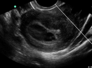
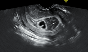
Now, this guy looks sick.  Legitimately sick.  Not just unsteady or a little too drunk to walk, he is GHOSTLY pale and unable to stand.  He’s got a pained grimace on his face and is about to pass out.  We got him back into his chair and someone went off to grab an extra bag of fluids and the ultrasound for me, since his heart rate had spiked up to 125.  After he was flat in his chair, I threw the ultrasound probe on his abdomen: RUQ was equivocal, but his LUQ had a nice huge stripe of free fluid.  He also had some new ecchymosis in his left lateral abdomen/LUQ.  Unfortunately I was in a little bit of panic mode and didn’t save his ultrasound images, but it was crystal clear to me what had happened: something had been bleeding for a while and was now causing a big problem.  So we rolled his chair into Room 9, plopped him onto a bed, and hit the Level I button when his manual systolic pressure came out at 85.  We pumped fluids in him and got blood to the bedside and into his veins right about the same time the wedge and chief showed up.  He stabilized enough for the scanner after initial resuscitation, and Trauma stood by in radiology to watch the images come up.

His spleen was pretty much ripped in two.  He didn’t appear to have any active extravasation (surprisingly, as far as I recall), and his pressure was improving though his heart rate wouldn’t go below 100 for more than a few seconds at a time.  I talked to him, I talked to his mother, and I talked to the Trauma folks.  We had enough time to get repeat blood work, discuss the impending surgery, and get him packaged up as stable as he was going to be before they whisked him away to Room 4 (or maybe 6, whichever).

He recovered uneventfully from his splenectomy, and was discharged from the hospital a few days later.  He had the best possible outcome given the particulars of his eventful time in the ER.  But I kept asking myself a handful of questions over the following days that I still think about from time to time.  What if we had walked him earlier, well before his hemorrhage was on the border of Class II and Class III?  Could he have gone home and died of hemorrhagic shock from a missed spleen injury?  What if I had scanned him earlier and he had only had a tiny contained rupture with no extravasation?  Would he have ended up in OR 4 anyway but gone there from the floor or PCU instead of from the ED?  How should I have proceeded differently?

After kicking myself for a couple of days, I talked with the attending who had been on with me at the time all of the kerfuffle went down, and felt much better about my decision-making process.  I distinctly remember him having no torso trauma or pain on my first assessment.  Man scanning this young, otherwise healthy patient was not indicated.  It was not contraindicated, but would have seemed superfluous and a misuse of resources based on his initial presentation.  As a side note, which was honestly extremely relieving to me, Trauma wasn’t all that critical of my decision-making process and was just glad we caught it before it got any worse.

In doing a little bit of background research, it appears that delayed spleen rupture is a not-entirely-unheard-of entity, but is debated in some surgical literature as a term coined as an alternative way to describe a missed initial diagnosis.  Regardless of what it’s called, it does happen.  Though, when it does, it’s usually after at least a couple of days (sometimes even a week), not just a few hours.  The literature focuses more on whether it’s a legitimate problem than how to manage, as the management is no different from a normal spleen rupture.  Operative intervention is the usual course, though some small lacerations/subcapsular hematomas are electively managed with observation first, especially in high-operative-risk patients.

My experience gave me 3 lessons to take away.

First: Young, healthy, adult patients can trick you just like children, with vitals not markedly abnormal until a big problem is present.  So you need to reassess them.  I know I’m as guilty as the next person of not always reassessing as thoroughly as I’d like, especially on a busy shift, but I’m also much more aware of who will need me to do a little more work before I can bless their departure.

Second: Drunk (or otherwise intoxicated patients) can trick you and hide serious problems.  Be aware of this going forward, because it doesn’t mean you need to man scan everyone, it just means you need to keep your mind open to injuries that aren’t initially apparent.

Third: Adjusting the plan and course is entirely okay, and in some ways it’s what EM is built for.  It’s why we get gaits on traumas with leg pain.  It’s why we walk our drunks and PO challenge our vomiting patients.  And it’s something we have to keep in mind because we’ve all seen patients who went from apparently fine to nearly-dead in what seemed like a single instant.

Prolonged QT

58 yo F presents to the ED for cough, chest pain, and fatigue for 1wk. She has sharp, atypical sounding chest pain. But she is 58 and has risk factors: HTN, HLD, 0.5ppd smoker x48yrs. Better get an EKG.

Prolonged QT EKG

Initial EKG

Awesome, no STEMI. Done with EKG right? Hope you didn’t miss that really long QT interval.

First, how do we measure the QT?

QTc image
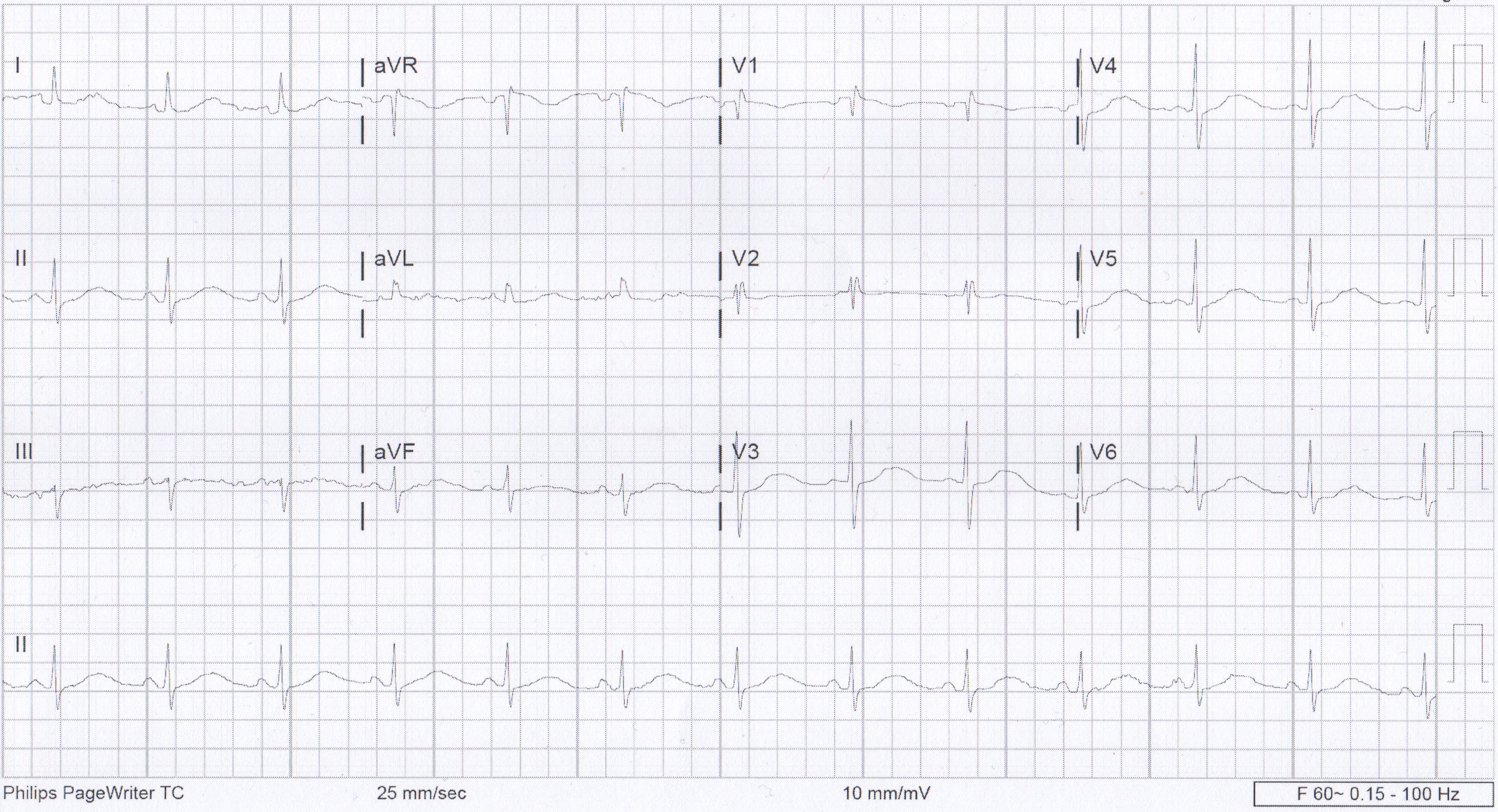
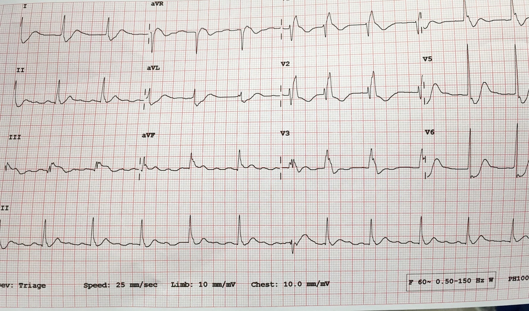
We usually talk about QTc rather than just the QT. This is because the QT interval varies depending on the HR. Using a correction equation standardizes the interval so it can be interpreted regardless of the HR. The EKG computer does give a calculation of the QTc, because we are obviously not hand calculating this on every patient. You should compare what the computer calculates to your gestalt when you review the EKG. If there are any concerns or discrepancies, you should hand calculate the QTc. MDCalc has an easy to use calculator. Above is the Bazett’s formula, which seems to be the most commonly used. Other formulas do exist. QTc is considered prolonged if > 440ms in men or > 460ms in women.

Next, why do we care?

ecg_hypokalaemia_torsades

This is the start of Polymorphic VT. There are several things that can cause this rhythm. Long QT is one possible cause. When Polymorphic VT is caused by a prolonged QT we give it a special name, torsades de pointes. The mechanism behind this is demonstrated on the above EKG. As the QT interval becomes more prolonged, there is a higher chance for an R-wave to hit on just the right part of the T-wave and cause this rhythm. QTc > 500ms seems be associated with higher risk.

So what causes prolonged QT?

Many things can cause prolonged QT. The most common etiologies are electrolyte abnormalities and drugs. Hypokalemia, hypomagnesemia, and hypocalcemia are well known to cause prolonged QT. Potassium and calcium are included on the CMP but don’t forget about magnesium. Drugs are also a big cause of prolonged QT. The list of drugs is long. Probably too long to memorize. However, there are some common medications and medication classes that you should know. The big classes are, Antiarrhythmics (like Amiodarone), Antihistimines (like Diphenhydramine), Macrolides (like Erythromycin), Antipsychotics (like Haloperidol), and TCAs (like Amitriptyline). This is not a complete list, just some highlights. If you really want to know if a specific medication is associated with prolonged QT, www.crediblemeds.org is a good source.

Other causes worth mentioning are structural heart disease, cardiac ischemia, and stroke. Some people do have Congenital Long QT Syndrome, but this should not be the leading diagnosis in the ED. Also those people are still at high risk for developing Polymorphic VT.

How did our patient do?

Her medications were reviewed and she was not on any of the most common offenders. Then routine labs came back. Unremarkable, except for K of 2.9! Repeat EKG after potassium repletion.

Normal EKG

EKG after K repletion


References:

  • www.uptodate.com
  • http://lifeinthefastlane.com/ecg-library/basics/qt_interval/
  • http://hqmeded-ecg.blogspot.com/2013/10/polymorphic-ventricular-tachycardia.html
  • bjsm.bmj.com/content/43/9/657/F3.large.jpgamp

Diabetes Insipidus in Intracranial Injury/Trauma

During a string of nights, I had two separate patients that despite having different injuries presented me with an interesting question. The first was a man in his 50s with a large intracranial hemorrhage, mass effect and the beginning of herniation; the second was a young male that was the victim of a GSW to the head. However, despite the different etiologies of their injuries, they both presented to Room 9 literally yelling for water. One became so combative in his demands for water, he had to be restrained.

This got me thinking; in the setting of very serious injuries, why is the only thing that concerns these men oral hydration?

The most concise information I found comes from Life in the Fast Lane. While it is referring to TBIs, the pathology relates to both of my patients as well. Thirst is controlled mostly by ADH released by the hypothalamus and transported to the posterior pituitary. Any disruption in this production chain can decrease ADH, leading to central diabetes insipidus.

In the setting of trauma, this can be caused by direct damage to the hypothalamus or posterior pituitary, disruption in their vascular supply or increased intracranial pressure/herniation that can compress these structures. Whether by a large hemorrhage or direct trauma like a GSW, intracranial injury can damage the ADH supply, leading to diabetes insipidus and the extreme thirst felt by these patients. Endocrinopathies have been associated with 30-50% of TBIs with the most common disorder being diabetes insipidus.


Just another overdose…..right?

20 yo M with unknown PMH comes in to room 9 with AMS and tachycardia to the 180s. Per EMS, he had been found down in his apartment, with crack cocaine pipes and other drug paraphernalia around him. He was found to be tachy as mentioned, as well as febrile with a temp of 103 axillary.

When he arrived in room 9, his HR was still in the 170s-180s. Blood pressure normal. He was pale, diaphoretic, and looked sick. He was speaking inappropriate words and would localize pain, GCS 12. Pupils dilated and briskly reactive. Rectal temp 104.1. CXR normal. Started IVF bolus and placed ice packs to the groin and axillae. Also gave Ativan as this was likely a stimulant overdose.

First EKG showed SVT at 180 BPM. After 2L of crystalloid and ativan, a repeat EKG showed sinus tachy at 140. The pt’s mental status was unchanged. The iStat showed a lactate of 13.

The plan was to place a rectal probe and monitor his temp, give him more fluids and Ativan prn, and re-assess later. I thought this was 100% an overdose. No problem.

About 2 hours and who knows how many room 9s later, I go to review his labs. I haven’t heard anything from nursing other than him continuously pulling out his rectal thermometer probe, so all must be well…Turns out he has a WBC of 44,000. Lactate has trended down, but he is still febrile to 102. This is when it hits me that maybe the guy who I’ve been treating for stimulant overdose is actually septic? His CXR and UA were normal, but maybe he has meningitis or encephalitis and that’s the reason for his mental status? Maybe I’m now 2 hours late with ABx?

I suppress the awful feeling in my stomach and go re-evaluate the pt. His mental status is unchanged from when I saw him in room 9. At least now his HR is in the low 100s. Given his mental status and tenuous vital signs, I know this patient is going to have to come in to the MICU. He’s going to need a head CT and an LP to rule out meningitis. I gave him antibiotics and called MICU. They evaluated the patient, and they agreed.

I chart checked the patient the next day. His LP was normal. His mental status improved overnight and he was transferred to the floor. Turns out this actually was likely all tox-related, but I thought it was a good learning point nonetheless. Sometimes it’s convenient to go down the path you’re led to by EMS or by nursing. Not only is it easy, but it’s usually the right path anyway. The stroke buzzer goes off and you immediately get your quick assessment over with so the patient can go to CT and stroke can do their thing. EMS tells you they found the patient in a house with drug paraphernalia, so you run with that.

But it’s important to keep the differential wide open when you first see a patient. At least consider less likely and less obvious possibilities. At some point, you’ll catch something that you otherwise would have missed until it was too late.

A Mysterious Death in a 21 yo Healthy White Female, and the Larson Maneuver

My wife is at work at a hand surgery pre-op care clinic. It is her birthday. In walks a middle-aged male who is about to have tendon reconstruction after he sustained a crush trauma, and is excited about the possibility of returning to work. He is slightly abrasive and somber, despite the prospect of receiving the surgery.

My wife asks him if he is concerned about the procedure, asks if he is feeling well, asks if there is anything she can do to help. With a quiet manner he states all is well. To make discussion my wife states today is her birthday and she is excited to celebrate when she gets home. He smiles and states today is also his daughter’s birthday. She was born the same year as my wife. He states today is always a rough day for him because she passed away in an emergency room 7 years ago at 21 years of age.

We can never fully understand where those we treat are coming from, their life experiences, or what their home situations, thoughts, fears, dreams, and worries entail; and this was a reminder for me. He stated that his daughter was healthy,woke up without issue on that day, but later developed difficulty breathing and wheezing. She was diagnosed with an acute asthma attack. She had one episode in the distant past but was not on any routine medications. She was given albuterol and experienced a negative reaction to the albuterol and completely stopped breathing. She was brain dead by the time she was intubated. They withdrew care in the emergency department.

After my wife shared this with me, I searched to find any case reports of paradoxical reactions to albuterol. Below are three related cases, however bronchospasm becoming worse with beta-blockers is exceptionally rare.

Case reports of paradoxical bronchospasm to inhaled beta agonists:

What I think more probable, and possibly related to the above case reports, is acute laryngospasm. The albuterol she received may have further irritated her vocal cords potentially worsening, rather than relieving her vocal cord dysfunction. Both Resus.me and LITFL (Life In The Fast Lane) have very useful articles describing management (see below for links). Here is a brief synopsis:

Laryngospasm

What is it?: a potentially life-threatening closure of the vocal chords (can occur spontaneously). Often misdiagnosed as asthma—especially exercise-induced asthma (more common in white females).

How to diagnose (and differentiate from asthma):

  • Stridorous sounds are usually loudest over the anterior neck, beware wheezing sounds transmit throughout the lungs
  • Typically, albuterol has minimal to no beneficial effect.
  • Subjectively more difficulty on inspiration than expiration

   Clues in history: recent exercise, GERD, ENT procedures, or extubation

   Common causes & some that are not-so common:

  • Post extubation
  • Exercise
  • GERD
  • Medications (e.g., (1) ketamine sedation, incidence 1-2 %; (2) versed (very rarely), which can be reversed with flumazenil)
  • Near drowning/ aspiration
  • Inhalants (smoke, ammonia, dust, cleaning chemicals)
  • Related to anxiety
  • Strychnine (plant based poison, sometimes used as a pesticide for birds and rodents, also the poison reportedly used to kill Alexander the Great in 323 BC)

Treatment of laryngospasm:

Initially:

  1. Jaw thrust with Larson Maneuver
  2. CPAP/ NIPPV
  3. Heliox might be helpful if available, (also topical lidocaine can be applied to larynx if available)

If conservative measures fail:

  1. Low dose propofol (0.1 mg/kg) ~ give 10 mg
  2. Low dose succinylcholine (AKA: suxamethonium) 0.1-0.5 mg/kg IV
  3. All else fails: intubation with succinylcholine 1.5 mg/kg IV
    • If no IV access, then succinylcholine IM (3-4 mg/kg). Experts advocate IM injection into the tongue.
    • Perform chest thrust maneuver immediately preceding intubation to open the vocal cords and allow passage of the ET tube.
    • Monitor for negative pressure pulmonary edema—(from patient pulling hard against closed glottis in the setting of acute asphyxia).

Flow chart from Resus.me

Larygospasm_flow_high_res

What is the Larson Maneuver? (Published 1998 in Anesthesiology)

It is a manipulation jaw thrust technique targeted at the ‘Larson’s point‘, AKA: laryngospasm notch.

  • Place middle finger of each hand in the laryngospasm notch, located behind the lobule of each ear, between ascending ramus of the mandible and the mastoid process.
  • Press very firmly inward toward the base of the skull with both fingers
  • At the same time lift the mandible at a right angle to the plane of the body (perform jaw thrust).

Reportedly will convert laryngospasm within one or two breath cycles to laryngeal stridor, and in after a couple more breath cycles, to unobstructed respirations. As proposed by Larson, it is likely that the painful stimulus relaxes the vocal cords by way of either the parasympathetic or sympathetic nervous systems through the glossopharyngeal nerve.

Diagram from LITFL

Larson_man

References:

  1. Resus.Me: http://resus.me/laryngospasm-after-ketamine/
  2. LITFL (Life In The Fast Lane): http://lifeinthefastlane.com/ccc/laryngospasm/
  3. UpToDate: https://www.uptodate.com/contents/paradoxical-vocal-fold-motion?source=machineLearning&search=Laryngospasm&selectedTitle=1~150&sectionRank=1&anchor=H3#H3
  4. Larson, Philip, MD. Laryngospasm-The Best Treatment. Anesthesiology. 1998. http://anesthesiology.pubs.asahq.org/article.aspx?articleid=1947036
  5. Paradoxical bronchospasm: a potentially life threatening adverse effect of albuterol. South Med J. 2006 Mar;99(3):288-9. http://www.ncbi.nlm.nih.gov/pubmed/16553105
  6. Paradoxical response to levalbuterol. J Am Osteopath Assoc. 2008 Apr;108(4):211-3. http://www.ncbi.nlm.nih.gov/pubmed/18443029
  7. Paradoxical reaction to salbutamol in an asthma patient. Pneumologia. 2012. Jul-Sep;61(3):171-4. http://www.ncbi.nlm.nih.gov/pubmed/23173379

Hypotension

Late 70s year old female with chief complaint of dizziness and fatigue. Patient has a medical history of HTN and recent cataract surgery.  In triage patient was hypotensive with BP of 70\35 and Bradycardic with a heart rate ranging from 55-60, O2 sats 100% on room air and afebrile.

I found the patient to be lethargic. Otherwise her exam was unremarkable with no focal neurologic deficits, cardiac and pulmonary exam unremarkable, and no abdominal pain. Her husband was in the room and states that they were on their way to their grandson’s high school graduation and she began complaining of feeling dizzy and she started to become lethargic. She had not been sick recently and before this morning she was completely at her baseline. To note yesterday her blood pressure was 180/95 when she checked at home so she typically runs high. No new changes to her medications which consisted of metoprolol Succinate 100mg QD and a Baby Aspirin.

As I talked to the husband he went on to explain that she had recently had cataract surgery on her left eye and that this morning she had a follow up appointment with her ophthalmologist. While at the ophthalmologist appointment the doctor said that the pressure in her eye was high and she received some drops in her eye to bring the pressure down but the husband could not remember the name of the drops.

So I started by getting CBC, CMP, TSH, Urine, Chest xray, EKG, Troponin and a head CT to complete my little old lady AMS workup. Obviously the differential diagnosis for AMS in the elderly is vast so I was considering a lot of different possibilities.  While I waited for her results to come back I called her Ophthalmologist that she had seen that morning to see what drops she had received. Ends up she got Alphagan which is an alpha agonist, Trusopt which is a carbonic anhydrase inhibitor, and 3 drops of Timolol. I discussed with him the possibility of the Timolol on top of her morning metoprolol 100mg as potentially causing her hypotension and bradycardia. He stated he had never seen that happen in his 16 yrs of practice but it is theoretically possible. A quick Uptodate search confirmed that hypotension can occur in as many as 10% of patients using Timolol eye drops which to me was a surprisingly high number.

So I’ll go ahead and cut to the good stuff. All of her labs and imaging returned unremarkable and her EKG just showed sinus bradycardia with a rate of 57.  Ultimately she got 2L of Normal Saline and we watched her for about 5 hrs. Throughout her stay her blood pressure steadily increased and at the time of her discharge she was 135/86 with a heart rate of 74 and she was back to her baseline and much more awake.

I thought this was an interesting case as it seems relatively rare for topical eye drops to result in systemic side effects however in the right patient population it can result in severe side effects. A quick literature search brought up multiple case reports of patient’s having symptomatic bradycardia and even syncope resulting from Timolol use.

So definitely something to keep in mind if you have a elderly or frail patient with acute angle glaucoma who is already on beta blocker therapy. Maybe trying other drops first instead of Timolol or at least be sure to make the patient aware of the possibility of side effects including hypotension, bradycardia, fatigue, and even syncope so they know what to watch out for.

Sick PEs

We had a very ill patient recently. She was found down upstairs visiting her family member. She was calmly altered, not agitated but was in mild distress. Consciousness fluctuated. Tachycardic and hypertensive initially, then had more labile BP and some hypotension.

We had to intubate her due to poor MS and clinical condition. She coded in CT, we placed a central line on the CT table between her noncontrast head CT and her CT chest. We pushed an amp of Epi and ran out of the room for the CT chest. We were worried about dissection and PE in equal amounts. We could not get good cardiac windows on bedside Echo in room 9 prior to the CT.

She continued to intermittently lose her pulse and drop her BP. We confirmed bilat PEs on the CT when we saw NO contrast left her right ventricle. The CT tech noticed first and became worried the patient had no cardiac output (ie pulseless).

We rushed the patient back to room 9 and gave a tPA bolus (50mg) followed by infusion of 40mg. She was on pressors and heparin and improving. Dr Smith accepted her to Jewish for possible EKOS or even ECMO if needed. On arrival to Jewish a few hours later she coded and died.

I was surprised when I found out she had died. Her O2 sat was improving, HR was decreasing, blood pressure was stable (though dependent on pressors). She received a large amount of crystalloid IV which according to some data might not have been optimal management. She also had the following ECG:

FullSizeRender

I think she was infarcting her myocardium. She likely had pulmonary infarction considering her poor oxygenation. She had coded a few times. She had a lot of strikes against her. Her BEST SHOT was going to a place with catheter assisted treatment for PE and ECMO if needed.

I am posting the case to let everyone know:

  1. How to manage sick PE patients (see post below)
  2. To use tPA in massive and in many cases of submassive PE
  3. TRANSFER sick PE patients to Jewish for EKOS/ECMO
  4. The decision to diagnose PE with RV strain on BEDSIDE Echo with no formal Radiologic testing will depend on your attending

This post from EmCrit / PulmCrit is a beautiful summary with potential dogmalysis related to PE management (see take home points below but do read the post).

In addition, here is a nice review article on catheter-based reperfusion treatment for PE with nice references for further reading.

Take home points from the Emcrit post:

  • The only evidence-based intervention that seems to improve mortality in massive PE is thrombolysis.   The primary goal of therapy should be administration of thrombolysis as soon as possible to patients without contraindication.
  • Consider early stabilization of blood pressure using a norepinephrine infusion, administered peripherally if necessary.
  • Volume administration may facilitate dilation of the right ventricle and hemodynamic deterioration.
  • Intubation is very hazardous and should be avoided if possible.   Patients die from cardiovascular collapse, and intubation may worsen this.
  • For a coding PE patient consider 50mg alteplase bolus as well as an infusion of epinephrine.  Patients can do well despite requiring CPR and high dose vasopressor infusions.

Phrenic Nerve Paralysis after intrascalene nerve block

This was covered in the opening of the February EMRAP however not something that I was at all familiar with.  Here are the basics and something that is probably rare but worth knowing about:

 In the episode they presented a case of  70 year old female that was brought in with a complaint of shortness of breath. RR = 28 and SA02 – 88%. The patient presented three hours after a right shoulder arthroscopy.  

ECG : normal

CXR: elevation of the right hemidiaphragm

 Dx:  paralysis of right hemidiaphram after intrascalene nerve block

There are two major complications associated with intrascalene nerve blocks: 1. pneumothorax

2. unilateral phrenic nerve paralysis.

The patient likely had a transient phrenic nerve dysfunction causing unilateral diaphragmatic paralysis.  Younger patients can compensate, older patients with co-morbid conditions may not be able to tolerate this as only one lung is effectively ventilating.

The patient in this case was managed with supplemental 02 until the buvipicane wore off.  Some patients with underlying lung disease and this complication may require BiPAP/CPAP or intubation.  

Things that I took away:

  •  be aware of this procedure and this complication
  • this may be done for patients with same days surgeries to the upper extremity and this is important history to have from Pt or family.
  • It may be missed prior to d/c.  This was an example of one that was missed by Anesthesia prior to d/c of the patient. 
  • May look clinically like PE (tachypnea, tachycardia, post-op patient), however,  history will help make the dx as well as CXR.


A little more about the block:

Interscalene nerve block is typically performed to provide analgesia for upper extremity surgeries and may or may not be combined with mild general anesthesia.

 Example of Surgeries this may be used for:

-Shoulder surgery, such as rotator cuff repair, acromioplasty, hemiarthroplasty, and total shoulder replacement

– Humerus fracture

Dr Huecker Stayin’ Healthy

As I was catching up on my weekly EM/pharmacology blogs, I stumbled across an article featuring our very own Dr Martin Huecker on the Academic Life in Emergency Medicine (ALiEM) website.  It is a great article about staying healthy and balancing life in EM. I’ve attached the link for your viewing pleasure.

https://www.aliem.com/2016/martin-huecker-healthy-in-em/

Spice/Heroin Reactions

So I’ve had to encounter my two sickest patients in the holding area within the past 2 weeks or so. One was a reminder from intern year while looking through spice charts, while the other was an actual patient that I had 3 days ago.

We always tend to take the “Tank” patients lightly and overlook them sometimes. I just want to use this as a warning (especially to the interns) that sick patients can also use heroin/ETOH/Spice, so pick up on the small interactions that don’t go right. I’ll try to present these starting with how their chief complaint lead to the final diagnosis.

Patient 1 (intern year)

  • middle aged male
  • CC: Spice OD, Nausea/Vomiting
  • Final Diagnosis: Subarachnoid Hemorrhage
  • Time to Diagnosis: 7 hours

So this guy presented as a spice reaction. This was before spice became as widely spread as it is now, and no one knew what to expect symptomatically (not that there is ANYTHING that is characteristic to spice anyways).

General story from talking with this guy is that he used spice for the first time that night. No significant past medical history. He was on a first date where everything had been going well. He had borrowed some spice from his friend where he used back at his place after dinner. Soon after he started having nausea and projectile vomiting and was acting ‘goofy.’ At that his date called EMS and the date ended.

Exam:

  • Gen: Fully A&O, slightly odd in that he seems incredibly happy to be here
  • CV: RRR
  • Pulm: CTAB
  • ABD: NT/ND
  • Neuro: CN II-XII intact, motor intact, sensation intact, ambulates without difficulty to bathroom

This man was like most of our intoxicated patients–a sober re-evaluation. At approximately 2 hours he was still vomiting in the ED, so the medical workup was initiated. Due to his odd behavior with vomiting, we got a CMP/CBC/Tox & CT Head. The night continued busy and I almost forgot about him as I waited for results. Ultimately he never got his CT Head due to being uncooperative but I wasn’t told until hours later. He ended up getting Geodon/Ativan in the ED but instead of calming him down he became more agitated and was no longer oriented. Ultimately getting rolled into room9 to be intubated prior to CT and the final diagnosis was made.

Certain forms of spice that lead to agitation also lead to spikes in blood pressure, and there are a few case reports of significant hypertension occurring after spice use. This guy had the unfortunate case of rupturing an aneurysm after using spice likely from a BP spike. I’m honestly not sure if the outcome would have been any different had I reached the diagnosis sooner — he got repetitive Head CTs and ultimately an EVD on hospital day 3. I didn’t really take him seriously even after I ordered a lab workup. This really changed my perspective on patients being held for intoxication. He spent 1.5 months in the hospital (1 month intubated) before being discharged to rehab.

Patient 2 

  • Middle aged white female
  • CC: Heroin OD got Narcan
  • Final Diagnosis: Cardiogenic Shock
  • Time to Diagnosis: 3.5 hours

This case I handled a bit better (I’d hope after 2 years). Story I could get is that this man had a syncopal episode. Received Narcan PTA by EMS and woke up. In the ED the patient adamantly denies heroin use–states he simply passed out. Luckily I got to him before EMS left, and EMS confirmed reports of bystanders stating opiate use.

Exam:

  • Vitals: HR 120, RR 16, O2 96%, BP 80/45, T 98.0
  • Gen: Fully A&O, drowsy
  • CV: tachycardia
  • Pulm: CTAB
  • ABD: NT/ND
  • Neuro: CN II-XII intact, motor intact, sensation intact, ambulates without difficulty to bathroom

My initial thought was that he may need some more narcan or that he received a longer acting opiate. The tachycardia was a wild card and didn’t make much sense with the picture. He remained afebrile and temp recheck, so I wasn’t thinking sepsis much at that time. At this point due to the tachycardia not making sense I ordered labs (and a tox for co-ingestants) and thought his BP/HR would improve with fluids.

I reassessed him after bolus #1 and #2 and neither HR or BP improved. Labs returned with an elevated WBC at 19.6. Opiates positive on top but otherwise were unremarkable. EKG sinus tachycardia. CXR and urine unremarkable. At this point even though I had no fever or source I felt compelled to initiate a septic workup and Lactate returned at 7.9.

I was starting to get lost as why this guy was so unresponsive to fluids and O’Brien and I threw the USN to bedside at this point. Turns out he was in acute systolic failure with an ejection fraction of 11%. No history of CHF and also no signs of volume overload on exam except very mild pulm edema. Troponin peaked at 0.5.

He was admitted by cardiology while they trended his status. He went to the cath lab on hospital day 3 with clean coronary arteries. Ejection fraction improved to 60% by time of discharge. Talking with the team today they are still uncertain of the cause.

These are two cases of sick patients being in the holding area. Hopefully, it serves to remind everyone that any patient can be sick.

Interesting Ultrasound

A late 20s F G4P3003 at approximately 6 weeks gestation by LMP presents with a chief complaint of vaginal bleeding. A few hours PTA, patient states she felt a “gush of blood” with some mild abdominal cramping. VSS. On exam, noted to have a moderate amount of vaginal bleeding per the os. On our bedside ultrasound we note what appears to be a viable IUP with cardiac activity.  However, the uterus appears septate, with half containing the IUP and the other half more hyperechoic/solid in nature. We were concerned for a possible subchorionic hemorrhage and consulted OB/GYN. Our bedside US image is below:

BS US

OB came down with their Cadillac ultrasound and confirmed our findings.  For comparison, here is their much clearer image:

OB US

For this patient, with this large of a subchorionic bleed, the likelihood of her carrying this pregnancy to term was low. They planned to have her follow up in clinic for a repeat ultrasound in 2 weeks to reassess viability. Per our OB colleagues, other things on the differential included a fibroid. However, as this patient had 3 very healthy and rambunctious boys at the bedside with her, OB commented that a fibroid that large would likely have resulted in infertility.

And from UptoDate:

“A subchorionic hemorrhage or hematoma is a risk factor for spontaneous abortion, particularly when it amounts to 25 percent or more of the volume of the gestational sac. A meta-analysis of seven comparative studies found that women having a subchorionic hematoma had a significantly increased risk of spontaneous abortion, compared to women without such findings (18 versus 9 percent; OR 2.18, 95% CI 1.29–3.68). The findings also are associated with an increased risk of placental abruption (4 versus 1 percent; OR 5.71, 95% CI 3.91–8.33) and preterm premature rupture of membranes (4 versus 2 percent; OR 1.64, 95% CI 1.22–2.21). The increased risks of preterm labor and stillbirth appeared to be dependent upon the presence of vaginal bleeding.

Pregnancy outcome associated with subchorionic hematoma also relates to location, with worse outcomes observed for retroplacental hematomas, compared to marginal hematomas. The location, rather than the size, of a subchorionic hematoma may be the most important predictor of pregnancy outcome. Evidence relating to the size of the hematoma and the risk of adverse outcomes is inconclusive.

The only management option for subchorionic hematoma is expectant. There is insufficient evidence regarding whether bed rest decreases the risk of pregnancy loss when a subchorionic hematoma is present. Some clinicians repeat an ultrasound in one to two weeks to confirm fetal viability and assess any change in size of the hematoma, primarily to provide reassurance to the patient. A subchorionic hematoma is not an indication to conduct a diagnostic evaluation for an acquired or inherited thrombophilia.”

Humanity

This is a commentary on this article by a resident reflecting on life, trauma, and death. The  resident’s self-reflection is a nice read and something with which we can all identify. But the “meta-reflection,” if you will, is something we do not usually get to see in our EM journals. We read one or two essays in each Annals, sometimes in Academic EM and in JEM. I tend to read these first or tear them out and hold them until I can read in a quiet room and really concentrate on them. And I do my own reflecting on the themes and ideas presented by that person.

The commentary article describes the difficulty in a self-analyzing essay. The schizophrenia and detachment involved in presenting your own thoughts in this way. Dr Ratzan reviews some of the (sadly few) accomplished physician writers and their themes. William Carlos Williams was a poet/writer and physician. Richard Selzer, a retired Yale surgeon, is still living and I hope still writing. His books are so rich and he is so talented a writer that I can hardly read more than 20 pages at a time. Brilliant physician writers are rare but essential to the humanity of our profession.

And emergency medicine in particular, with its intensity and ?necessary detachment, might need this humanity more than any other specialty. I recommend we all read something either in the medical humanities, or something seemingly unrelated to medicine. You might think the literature or the book of humor or the young adult futuristic death competition book is non-medical. But connecting, or reconnecting, with that part of yourself that is separate from the ED will make you a better doctor and healer.

It can become trite to say feelings are important, we need to have empathy, there is a human side to this job of ours, etc. And it is a difficult jump from reading a feel-good essay to the next day walking into the room of a patient with fibromyalgia and trying to channel that empathy you were just reading about. But trust me, if you make an effort to do this you will appreciate your patients. Good advice I read from a Brazilian shaman: “remember that the world does not revolve around you.” you have to really think about that to understand. Try to picture the world through literally through other people’s eyes, makes yourself and your troubles seem smaller.

The patient you are seeing in the ER is likely having the worst day of his month or year or even life. Try not to forget that. Happy Thanksgiving.