Cool Name, Not So Cool Results

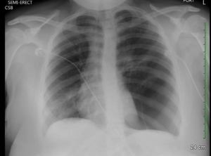
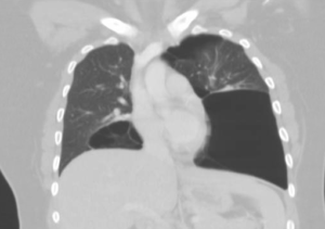
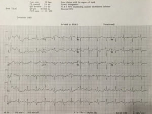
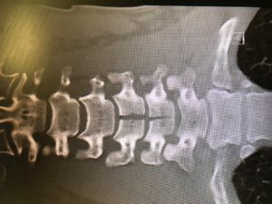
53yo WM rolls in, wee hours of the am.  C/o HA, onset around 4 days ago.  Wife says “I think he had a seizure in the middle of the night that night”    Def hit his head, has a goose egg middle of the forehead.   “Had some labs done at my primary yesterday, they called me and told me to come in”   No clue as to what labs those might be when asked.   Awesome.  Medical hx only significant for HTN and GERD.   No chronic meds.   Admits to drinking around a case of beer a day.   Probably explains the not so full history he provided.   Nothing else interesting on physical exam and normal vital signs.  So I send labs- CBC, Chem13, Mag, CT his head due to the possible seizure.

Annnnd Na 114, K 2.5, CL 75, Bicarb 32, BUN 12, Cr 1.2, Glucose 111, Ca 8.4.   CT normal.  ETOH and Tox NL.   Serum osmolality 259

So what’s going on here?   Hyponatremia and seizure- needs fixed right.   Normal saline or even hypertonic saline maybe?

Negative ghost rider

Lets look at this a little deeper.

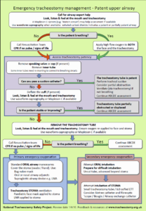
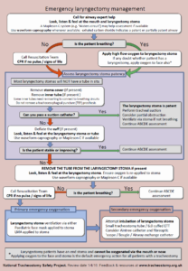
Start with the algorithm

Image result for hyponatremia algorithm

So we will start with the asymptomatic column.   Could argue for the severe side due to the seizure, but he is now 4 days out and asymptomatic.   Went the euvolemic side of things.   No signs of fluid overload clinically and didn’t look dry.  Actually nerded out and ordered urine osmolality.  88.    So where does that land us?

Beer Potomania syndrome.

WTH is that?  Great name.   But too much physiology for my brain.

When patients have poor protein and solute (food, electrolytes) intake, such as in chronic alcoholics, they can experience water intoxication with smaller-than-usual volumes of fluid. The kidneys need a certain amount of solute to facilitate free water clearance (the ability to clear excess fluid from the body). A lack of adequate solute results in a buildup of free water in the vascular system, leading to a dilutional hyponatremia.

Free water clearance is dependent on both solute excretion and the ability to dilute urine. Someone consuming an average diet will excrete 600 to 900 mOsm/d of solute. This osmolar load includes urea generated from protein (10 g of protein produces about 50 mOsm of urea), along with dietary sodium and potassium. The maximum capacity for urinary dilution is 50 mOsm/L. In a nutritionally sound person, a lot of fluid—about 20 L—would be required to overwhelm the body’s capacity for urinary dilution.

However, when you don’t eat, the body starts to break down tissue to create energy to survive. This catabolism creates 100 to 150 mOsm/d of urea, allowing you to continue to appropriately excrete a moderate amount of fluid in spite of poor solute intake … as long as you are not drinking excessive amounts of water.

Alcoholics get a moderate amount of their calories via beer consumption and do not experience this endogenous protein breakdown or its resultant low urea/solute level. With low solute intake, dramatically lower fluid intake (about 14 cans of beer) will overwhelm the kidneys’ ability to clear excess free water in the body.   

So, we see alcoholics all the time.  Why don’t we see this all the time?   When you think about it, a pretty low percentage of our EXI all star drinkers are beer drinkers.  And most case a day beer drinkers actually have a decent oral food intake.   Making this more rare than you originally might think.

So what do we do about it?  Classic example of ‘dont just do something stand there!’

Fluid restriction, fluid restriction, fluid restriction.

Back to my case.  Admit the patient to my hospitalist, who is half asleep and currently not as excited as I am about hyponatremic management.   Orders in for fluid restriction and serial chemistries.    A few minutes later, I hear my ER nurse arguing with the floor nurse during calling of report.   They are appalled that I am not giving normal saline, and even request hypertonic saline.  I politely pick up the phone and discuss the physiology and reason for my treatment plan.   We were on the same page by the end of the call.

Fast forward to a couple days later.  I come back for another shift, and am checking on my patients from the day before (quick aside, no matter where you are in your career always save the info for 3-4 of your sicker, more interesting patients and look them up on your next shift, by far one of the most high yield learning you can get.   And will help you adjust your practice as indicated).   I look up the serial sodium results.  114, then 119, 123, then………145.   Goooooooo!!!!  Assuming I am about to hear from the Kentucky Hammer due to causing central pontine myelinolysis.  I talk to the hospitalist- apparently the next shift nurse, after the one I talked to, got her way, and they got an order to blast the guy with normal saline.  Hence the huge jump.   Patient did have some transient AMS, but was at baseline and neuro intact once levels stabilized.  Thank god.

Anyway, interesting case.  Beer potomania.   cool name, not so cool results.   Literature states that central pontine myelinolysis happens in over 20% of these patients due to too rapid correction.  So before you pull the trigger on normal saline repletion take a second and scope out the algorithm above.  Sometimes the best thing you can do is nothing.

Video Links for Fiberoptic and Topicalization

As promised,
Several links with videos on the process of fiberoptic as well as topicalization. Lots of variations on strategies here, some of which of course are really more applicable for awake Anesthesia patients and may not fit our population but definitely helpful.
The Life in the Fast Lane has a good written summary as well. Hope these help. Email or talk with me for questions!


Good video of topicalization process for awake nasal and oral


Dr. Gallagher discussing the gist of fiberoptic intubation


Rich Levitan’s Video of Fiberoptic Intubation

https://www.youtube.com/watch?v=rljSPu7-vZA
20 min video from Anesthesiolgoist on Fiberoptic- decently covers range of topics

https://lifeinthefastlane.com/ccc/awake-intubation/
LIFTL summary with a couple different videos both on awake intubation and also fiberoptic

https://www.youtube.com/watch?v=c9pAQ3DUKVM
Dr. Ali Diba using aScope on Awake patient—uses spray as you go technique


Narrated Talk on Topicalization: They do both Nasal and Oral for an Oral Fiberoptic for some reason. Also used both viscous Lidocaine and Ointment then sprayed cords.


Video on the LMA MADgic

Critical Care Time

Good 3-5 minute read on Critical Care time complements of ALiEM- what qualifies, time frames, procedures, etc.

https://www.aliem.com/2017/07/charting-coding-critical-care-time/

Pantoprazole on Shortage

See below: The gist is, 40mg IV Pantoprazole (Protonix) on shortage until August or so. Consider using Pepcid instead.

Medical Staff,

Pantoprazole 40mg inj vials is on national backorder. The anticipated availability is in early August. To ensure continuity of patient care, Pharmacy, supported by P&T plan to substitute pantoprazole 40mg IV push daily to famotidine 20mg IV push BID for the indication of stress ulcer prophylaxis. And pantoprazole IV push to esomeprazole IV push for all other indications.

Pantoprazole 40mg inj vials will be reserved for existing PPI drip protocols.

The good news is that we do not have to adjust existing protocols or infusion pump library settings. Pharmacist will manually change the order from pantoprazole to alternative upon verification. We expect that this shortage will be relatively short.

Please call pharmacy with any questions or Michael Nnadi at 336-817-5265.

Narcissus: A Case Presentation

11 month old Chinese female brought with concern for two episodes of emesis after family dinner. Via interpreter phone the father explains that everyone in the family vomited after dinner at home. He states the adults all vomited once and feel fine now, but the child vomited twice and he wants to ensure she is well. He is concerned that she is sick from “the onions”. When asked to clarify he explains he made a noodle dish for dinner – every ingredient has been used previously for the same recipe except for “the onions”. He then holds up a Kroger bag of what initially appear to be green onions, with the typical long green stalks and a white bulb base – but these are covered in dirt. He explains that he found them growing in their backyard and thought he’d use them for dinner, but now suspects they have made everyone sick. A brief intradepartment search procured a gardener RN who readily identified the plants – daffodils, “they just haven’t flowered yet”.


Kentucky Regional Poison Control advised that all parts of the daffodil (genus Narcissus) cause self-limited gastrointestinal symptoms for approximately 3 hours after consumption, and that care is largely supportive. There are reports of massive consumption causing CNS symptoms in dogs, but similar presentations have never been reported in humans.


Take home points:

  1. Green onions and unflowered daffodils are quite similar in appearance – the classic onion odor and tearing following incision differentiates the two
  2. Daffodil toxidrome is self-limited, with predominantly gastrointestinal symptoms (nausea, vomiting, diarrhea). Supportive care with oral rehydration fluid is warranted in pediatric patients.

References:

  • National Capital Poison Center – http://www.poison.org/articles/2015-mar/daffodils

Limping toddler

Nothing crazy here, just some EM bread and butter. I’ve had a couple of these at Children’s, and each time (with two different attendings), I’ve been told that the adult EM residents seem to overlook this, or not have any idea that it’s a thing, which is kind of embarrassing.

The patient is a 22-month-old male who presents with difficulty walking. Mom states that the child was walking fine until this morning. Since then, he has not been wanting to put weight on his right leg. Mom does not recall any injury. The child is otherwise well, no signs/symptoms of illness, and he has no medical problems.

On exam, the child will not put weight on his right leg when forced to stand. The extremity is well perfused, and there are no signs of trauma. He has no point tenderness, so it is not clear where he is hurting, but does seem to have pain when the foot is grasped and rotated internally and externally.

Discussion: In the toddler with a possible lower extremity injury, it may be difficult to localize where the child is having pain. If there is a question, the entire extremity should be imaged (though you should try to localize the problem area if possible). In this case, we suspected a toddler’s fracture, so a 3 view tib/fib was obtained. This is an important learning point: many times, the fracture line will only be visible on the oblique view, so it is necessary to get 3 views. In this patient, the xray was negative (as were other films of the leg). We diagnosed the child with a toddler’s fracture, placed him in a short leg splint with stirrups, and discharged him with orthopedics follow up.

A toddler’s fracture is a spiral fracture of the distal tibia which usually occurs by the same mechanism as an adult spraining an ankle. Sometimes it is a clinical diagnosis, not visible on X-ray. There is debate in the literature about immobilization in this case; some say it is necessary, some say it’s not. The culture at Norton Children’s seems to be immobilization. Regardless, the child should follow up in 1 week for definitive diagnosis, either with repeat plain films, or possibly MRI or bone scan. If there is a visible fracture on plain films in the ER, the child should be placed in a short leg splint with knee immobilizer and follow up with orthopedics in 72 hours.

It’s my asthma

An interesting case from the Pediatric ED:

A mid teen female with a reported two year history of mild persistent asthma, presented with asthma exacerbation. She presented to an outside ED with two days of cough, wheezing and shortness of air which acutely worsened three hours prior to arrival. Upon initial presentation there, the patient was reported to be in moderate respiratory distress with increased work of  breathing, tachypnea, and an O2 saturation in the 80%’s on room air. Lung auscultation revealed diminished breath sounds on the left with faint wheezing at left and right apices. She was placed on 3L nasal cannula with normalization of her O2 sats. She received an hour long course of albuterol and dose of Solu-Medrol, with moderate improvement in her symptoms. She subsequently received a chest X-ray which was concerning for a large left sided bleb/cyst with mediastinal shift to the right. Patient was subsequently transferred to us.

Further HPI and PMHx revealed that patient was diagnosed with allergic rhinitis in her early teens and diagnosed with asthma two years ago. She reported no history of ever being evaluated with a chest x-ray prior to current presentation. Over the course of the past two years she had been prescribed 3 courses of steroids and used albuterol approximately 2x per week PRN. Patient denied any other medical problems or smoking history.

A CT scan of the chest confirmed presence of large left sided pulmonary cyst.

Patient was admitted and followed by both pediatric pulmonology and surgery. Inpatient workup included PFT’s and alpha-1-anti-trypsin (negative). She was eventually diagnosed with idiopathic giant bullous emphysema, otherwise known as vanishing lung syndrome. She later underwent bullectomy with improvement of her symptoms.

Bullous emphysema is often seen as a complication of COPD in adults, but rarely diagnosed in children with few case reports in the literature. Giant bullae can present asymptomatically, with progressive dyspnea, hypoxia or hemoptysis. Giant bullous emphysema is classified as occupying more than one third of a hemi-thorax, and is often initially mistaken as a pneumothorax on initial chest radiograph. As disease progresses, enlargement of the bullae fill with air and loss of lung function results as fibrous membranous surface of the bullae result in poor gas exchange. Bullae are at high risk for rupture and tension pneumothorax should be suspected if a patient develops worsening respiratory distress.

The learning point I took from this case is to step back and re-evaluate a patient’s presentation when things do not exactly add up, as in this case a teen with worsening asthma not responding to conventional treatment and little pcp workup in the past. This point is particularly exemplified in the pediatric population where we often see several low acuity cases each shift, often drifting into “auto pilot” mode, employing little critical thinking. Don’t forget, rare or more complex pathology may actually be responsible for the patients symptoms.

My Lesson on Anchoring

If I had to pick one case from intern year that truly taught me the importance of keeping a wide differential diagnosis, it would be my final Room 9 of the year. The buzzer went off, and as I made my way to the trauma bays, I was able to get a brief rundown from the attending. “Seizure, 40-sish male, no known history”. OK, this was something I could do. I began running everything I’d need to do through my head as I prepared for the patient. “ABC’s. Vitals. Fingerstick glucose. Ativan… Could be trauma, hypoglycemia, benzo or alcohol withdrawal…” As I was refining my differential, the patient came in. The patient was non-rhythmically jerking, was not responsive to voice or sternal rub. I noticed he was wearing dress pants and a collared shirt. He was breathing spontaneously and maintaining sats in the mid 90’s. Palpable pulses and good heart sounds. Glucose was in the 100’s. EMS said he had been found like this approximately fifteen minutes prior, and his clinical status hadn’t changed since then. No known medical history or medicines.  I called out for the nurse to draw up Ativan, as I said this I noticed the patient had urinated on himself. Everything in my mind pointed towards a seizure. I grabbed the otoscope to perform the secondary survey and pried open his eyes. That’s when I felt the rug come out from under me.

The patient had pinpoint pupils, one millimeter bilaterally. The attending and I immediately had the same thought, and as I opened my mouth I heard him say, “let’s get some narcan for this guy!” The narcan got administered quickly, and soon after the patient woke up agitated, but responsive. His family had driven to the ED soon after he arrived, so questioning him was difficult. We ended up taking him to privacy in an empty x-ray room, where he admitted to using heroin earlier in the day. Sure enough, his toxicology screen was positive for opiates. We counseled him, observed him in the ED to ensure he didn’t need another dose, and then discharged him home.

What struck me about this case was that while I had formulated a differential, I had done so after anchoring to a faulty premise. It taught me a valuable lesson in keeping my differential broad, and it’s a lesson I’ll carry to every patient encounter from here on out.

Serial ECGs

I had a fairly interesting patient back in November 2016 when I was at Jewish.  I had picked up a patient with chief complaint of chest pain after an attending handed me the EKG below.  The patient was being rolled back from triage to a room at the attending’s request. 42yo AAM with history of hypertension, hyperlipidemia, diabetes, sleep apnea and CAD status post CABG in 2008.  The patient was complaining of chest pain that initially started when he was sitting at home watching TV about an hour prior to arrival.  He described the pain as sub-sternal and radiating to his left arm. He had associated SOA, palpitations, and diaphoresis along with the pain. He was also nauseated since the onset of pain.

Pretty classic presentation here in a patient with previous heart disease.  His initial EKG recorded in triage is below:

Upon review, you can appreciate that there may be ST elevation in III.  Also, ST depressions are noted in the precordial leads along with ST changes elsewhere. My question to all of you based upon reviewing this EKG: Would you call this is a STEMI and would you activate the cath lab at this time?

The attending that I was working with at the time didn’t feel that we could definitively call this a STEMI based upon the first EKG.  We examined the patient and collected his history after he was brought back.  After labs were collected, the patient received nitro paste.  Despite the paste, he continued to have pain. After approximately 15 minutes of being in the back, the patient reported that his pain was worsening.  The decision at that time was made to get a repeat EKG.  The repeat EKG is below:


So now what are your thoughts? There is obvious ST elevation in the inferior leads.  There are also ST depressions in the precordial leads.  This EKG was taken 34 minutes after the first EKG which was performed in triage.  Through these two EKGs, you can appreciate the evolution of a STEMI.  This patient was emergently taken to the cath lab at that time and underwent left heart cath. He underwent an impella-assisted PCI to his SVG-PDA and was started on dual anti-platelet inhibitors. Following his PCI, he also had an episode of wide-complex rhythm which resolved after receiving amiodarone.

So this patient who was found to have a STEMI on his repeat EKG had presented with an initial EKG that was non-diagnostic for his condition. Did this affect his outcome in any measure? What can we learn from this?

According to a 2013 study conducted by Riley et. al, in a national sample of patients diagnosed as having STEMI (41,560 patients), 11.0% had an initial non-diagnostic ECG. Of those patients, 72.4% had a follow-up diagnostic ECG within 90 minutes of their initial ECG. There did not appear to be clinically meaningful differences in guidelines-based treatment or major inhospital outcomes between patients diagnosed as having STEMI on an initial ECG and those diagnosed on a follow-up ECG.

So, did the additional 30 minute delay in activating the cath lab affect this patient? Likely not. However, what this case reinforces is the importance of a repeat EKG as 11% of these patients likely will not have STEMI on their first EKG.


References:

Am Heart J. 2013 Jan;165(1):50-6. doi: 10.1016/j.ahj.2012.10.027. Epub 2012 Nov 21. Diagnostic time course, treatment, and in-hospital outcomes for patients with ST-segment elevation myocardial infarction presenting with nondiagnostic initial electrocardiogram: a report from the American Heart Association Mission: Lifeline program. Riley RF1, Newby LK, Don CW, Roe MT, Holmes DN, Gandhi SK, Kutcher MA, Herrington DM.

SIH – Spontaenous Intracranial Hypotension

Recent EMRAP podcast reminded me of a case from when I was a Medical Officer in the Navy.

One of my Marines, early twenties, with no medical problems presented for follow up in sick call after being seen in the emergency department for a headache.  Since it was the worst headache of his life, an LP was performed.  Both the LP and the CT head were negative. He was given standard headache treatment with “migraine cocktail” from what I remember, however, no significant relief.

He presented to clinic with complaint of ongoing headache that was only better if he laid completely flat with excruciating pain with sitting up.   He refused to do anything other than lay on the gurney in the treatment room.

His presentation was classic for post LP headache, however, he states that this was the same headache that he presented to the ED for the previous evening and was not changed by the LP.  He was adamant about this timeline and unchanging symptoms after LP. Physical exam was normal to include normal Neuro exam.

I subsequently called the Neurologist on call at Naval Hospital, who stated the likely diagnosis was a “spontaneous CSF leak” and recommended a blood patch.  I was quite confused as I had never heard of such and it seemed like a made up diagnosis to me at the time.   I did, however,  want to help my patient and for him to leave the clinic at some point that day.  I then called anesthesia who was agreeable to the blood patch given recent LP and current exam/symptoms.   The patch lead to resolution of his symptoms and he was able to stand up and walk without a headache.

This is the only case that I have seen, however, the recent EMRAP review leads me to believe that I might see another case while practicing Emergency Medicine.

SIH is caused by a spontaneous tear in the dura in the spine or elsewhere in the meninges and leads to intracranial hypotension from CSF leak.  Symptoms are incredibly similar to post LP headache given the pathophysiology on really differ in that CSF leak in an LP is iatrogenic and SIH is well, spontaneous.  Diagnosis can be by LP, which will demonstrate low opening pressure, or MRI of the Brain W/WO and Spine W/O which will demonstrate the leak.  The symptoms are similar regardless of the level at which the leak occurs so you may have scan the entire meninges to find it.

Treatment initially is caffeine and rest for mild-moderate headaches and lumbar blood patch for moderate-severe headaches.

Uptodate has a lengthy but throughouh algorithm for diagnosis and treatment.   If lumbar patch doesn’t work, a more targeted approach may need to be employed.

  • Take-home point – consider SIH when someone presents with post LP headache symptoms without having had one performed in the recent past.

Baffling Neurology Pathology Strikes Again…

…forever seeking the unsuspecting emergency medicine intern.

A previously healthy teenage male presented to the emergency department via emergency medical services with the complaint of weakness, sensory changes, and increasing difficulty of breathing.  Family reported the patient went outside to mow the grass approximately 10 hours prior to presentation.  After two passes in the yard with a push mower, the patient had sudden onset occipital headache, fatigue, parasthesias described as tingling in both lower extremities, and generalized weakness.  Patient reported these symptoms to parents and laid down to rest.  After an hour long nap, patient was encouraged to take a shower to see if symptoms improved.  During shower, patient became weaker, had one episode of vomiting and was no longer able to stand on legs.  The patient’s father reports patient was able to make small movements but unable to ambulate or push against resistance.  Emergency medical services were contacted at this point, however, after assessment, the symptoms were deemed related to anxiety and patient was not transported.  Over the course of the day, the patient’s weakness progressed to the complete inability to move legs, followed by inability to move arms, with continued paresthesias in all extremities. After patient demonstrated worsening respiratory distress, EMS was called again.  Parents denied history of asthma, prior wheeze, fevers, recent illness or trauma, recent travel or drug abuse.  Family reports patient went camping one week prior but denies tick exposure.


On exam, the patient had significant respiratory distress with poor air movement and was only able to answer questions with one word responses secondary to respiratory distress.  Neurological exam revealed 0/5 strength in all extremities, areflexic biceps and brachioradialis reflexes, areflexic patellar and Achilles reflexes, and downward going Babinski bilaterally.  Sensation to light touch was intact but diminished in all extremities.  There were no rashes or lesions on skin exam.


Initial differential included but was not limited to:

  • Organophosphate toxicity – although patient self-decontaminated earlier in the afternoon and did not have diarrhea, salivation, or lacrimation
  • Guillain Barre – although acute time course without report of recent URI or GI syndromes
  • Tick paralysis – although no known exposure or lesions identified on skin or scalp
  • Transverse Myletitis – although no personal history of recent illness, no family history of multiple sclerosis or other autoimmune disease
  • Spinal Cord Infarct – although patient and family deny trauma, recent surgery, or history of coagulation disorder
  • Conversion Disorder – although no anxiety or other psychiatric history

As concern for bronchospasm, the patient was given IM epinephrine and started on a hour long albuterol while history was being obtained.  Bedside ultrasound demonstrated grossly normal myocardial contractility without effusion, normal lung sliding, and a collapsible IVC.  Chest x-ray was unrevealing. After lack of improvement with initial intervention, second dose of epinephrine and fluid bolus begun.  Intial VBG demonstrated hypercapnea and patient was started on BiPAP.  Within minutes, patient showed improvement in respiratory status and appeared more comfortable.  Initial CMP, CBC, CRP, and ESR were unrevealing.   Lumbar puncture was performed after head CT revealed only an arachnoid cyst.  CSF studies demonstrated mildly elevated segs, mildly elevated glucose, negative gram stain and normal protein.  Foley catheter was placed after patient complained of bladder fullness (confirmed by bladder scan) with inability to void.  Neurology and Neurosurgery were consulted from the emergency department, who requested urgent MRI on admission.  Patient was admitted to PICU with ED diagnoses of acute flaccid paralysis and acute neuromuscular respiratory failure.   After admission, MRI was obtained and demonstrated (drum roll please) ischemia vs. infarct from approximately C2-T5 with predominance in the anterior horns.


The literature review of non-traumatic spinal cord infarction is as rare as the pathology itself in the pediatric population and predominately consists of case studies.  Causes typically include but are not limited to hypotension, vascular injury, thrombus, embolus or compression.  Considering many of these etiologies are already scarce in the pediatric population, finding cause proves to be a difficult task.   Pain in neck, back or legs, weakness, tingling, and numbness are commonly described symptoms in pediatric case studies and were present in the patient that presented to us.   Prognosis studies have been performed on primarily adult patients; one such study demonstrated >40% recovery in ambulation amongst the 37 patients that left the hospital wheelchair bound through aggressive physical therapy and rehabilitation programs.[1]   Poor prognostic factors include female sex, advanced age, severity of symptoms, and lack of improvement within 24 hours after infarction.[2]   It appears as though outcomes are primarily dependent on access to physical therapy and supportive care including psychological treatment.   Workup and treatment recommendations if suspicious of atraumatic spinal cord infarct include obtaining an MRI for definitive diagnosis, followed by autoimmune, hypercoagulable, and infectious evaluations. Treatment in the emergency department is mostly supportive and includes addressing any contributory conditions and the management of respiratory concerns.[3]


On follow up, I found that the patient required intubation after exhibiting respiratory decompensation after the MRI was obtained.  Initial treatment with high-dose steroids and IVIG were discontinued when infectious and immunologic work-ups were unrevealing.  The hospital course was complicated by neurogenic bowel and bladder, neuropathic pain, anxiety, intermittent autonomic instability with hypotension, pneumonia and a urinary tract infection.  The patient required tracheostomy and percutaneous gastric tube placement during his hospital stay.  The patient continued to exhibit flaccid paralysis of all four extremities and ventilator dependence on discharge to acute rehabilitation.


Per my own investigation, I was relieved to find that Frazier has both success managing these cases and many unique resources that may positively impact this patient’s course (if you are curious here is some video evidence https://www.youtube.com/watch?v=8o_w174rI3s).  Needless to say, my differential for motor weakness, particularly in pediatrics, will forever be haunted by this case.


[1] Robertson CE, Brown RD Jr, Wijdicks EF, et al. Recovery after spinal cord infarcts: long-term outcome in 115 patients.  Neurology. 2012; 78: 114–121.

[2]Cheshire WP, Santos CC, Massey EW, Howard JF Jr .  Spinal cord infarction: etiology and outcome.  Neurology. 1996;47(2):321.

[3] Spencer, Sandra P. MD; Brock, Timothy D. MD; Matthews, Rebecca R. MD; Stevens, Wendy K. MD.  Three Unique Presentations of Atraumatic Spinal Cord Infarction in the Pediatric Emergency Department Pediatric Emergency Care. 30(5):354-357, May 2014.

Snakes and ladders

Last month I had an interesting Room9 for visual purposes. The story per EMS was jumbled, as it can be from time to time. All we knew was that the patient was a middle aged man who either fell off a roof or jumped off a ladder into a foot and half of water. …agreed. Details were otherwise unavailable. We manage repercussions of injuries, not the causes.

The patent came in intubated, wet, hemodynamically stable. The patient had ketamine en route but was still active with GCS 6T. He required more than your typical sedation to be amenable for the CT scanner. The physical exam showed no motor activity in lower extremities, including to painful stimuli. We noted no step-offs or abrasions. The patient had no signs of trauma other than the motor weakness.He moved his upper extremities and needed restraints due to lack of response to sedation. CT images are below. Most of these images mirror almost exact images from Dr. Ferguson’s lecture on spine fractures, thus I thought it would be good to go over.

As you can see the patient had significant fractures of his cervical spine. Talking with Neurosurgery, the burst fracture is more common in lower thoracic and upper lumbar spine and only is seen in cervical spines to this degree under severe axial load injuries, such as going head first from a significant height (especially when the posterior column has a vertical fracture as seen above).

I’ve always wondered the significance of doing spinal check during the physical exam prior to CT, when they are already getting “manned”. I often feel that if there is real pathology (ie. unstable fractures), won’t palpating (and deep palpation on obese patients) worsen the fracture and theoretically cause neurological issues. I haven’t found much to substantiate that, but it seems to make intuitive sense; I am open to any other opinions / suggestions.

Does anyone want to comment on the type of fractures noted, stable vs unstable (refer to Ferguson’s lecture)?

Are teardrop fractures stable?

Is there any significant retropulsion?

Could you hypothesize flexion vs extension injury?


Answer: Unstable teardrop fracture as well as a burst fracture, and borderline chance fracture (not typical for this cervical spine location).


For further spine related information please look at the links below or Dr. Ferguson’s spine lecture. Ferguson’s is a great source covering the importance of stable and unstable fractures, and a great lecture for interns to go over, especially early on in the year.

Here is a good podcast from Scott W. on less traumatic c-spine injuries:

EMCrit 63 – A Pain in the Neck – C-Spine Imaging and Clearance

And some other good sources:

http://www.aafp.org/afp/1999/0115/p331.html

http://www.paems.org/pdfs/online-ce/Evaluation-and-management-of-acute-cervical-spine-trauma.pdf