It’s my asthma

An interesting case from the Pediatric ED:

A mid teen female with a reported two year history of mild persistent asthma, presented with asthma exacerbation. She presented to an outside ED with two days of cough, wheezing and shortness of air which acutely worsened three hours prior to arrival. Upon initial presentation there, the patient was reported to be in moderate respiratory distress with increased work of  breathing, tachypnea, and an O2 saturation in the 80%’s on room air. Lung auscultation revealed diminished breath sounds on the left with faint wheezing at left and right apices. She was placed on 3L nasal cannula with normalization of her O2 sats. She received an hour long course of albuterol and dose of Solu-Medrol, with moderate improvement in her symptoms. She subsequently received a chest X-ray which was concerning for a large left sided bleb/cyst with mediastinal shift to the right. Patient was subsequently transferred to us.

Further HPI and PMHx revealed that patient was diagnosed with allergic rhinitis in her early teens and diagnosed with asthma two years ago. She reported no history of ever being evaluated with a chest x-ray prior to current presentation. Over the course of the past two years she had been prescribed 3 courses of steroids and used albuterol approximately 2x per week PRN. Patient denied any other medical problems or smoking history.

A CT scan of the chest confirmed presence of large left sided pulmonary cyst.

Patient was admitted and followed by both pediatric pulmonology and surgery. Inpatient workup included PFT’s and alpha-1-anti-trypsin (negative). She was eventually diagnosed with idiopathic giant bullous emphysema, otherwise known as vanishing lung syndrome. She later underwent bullectomy with improvement of her symptoms.

Bullous emphysema is often seen as a complication of COPD in adults, but rarely diagnosed in children with few case reports in the literature. Giant bullae can present asymptomatically, with progressive dyspnea, hypoxia or hemoptysis. Giant bullous emphysema is classified as occupying more than one third of a hemi-thorax, and is often initially mistaken as a pneumothorax on initial chest radiograph. As disease progresses, enlargement of the bullae fill with air and loss of lung function results as fibrous membranous surface of the bullae result in poor gas exchange. Bullae are at high risk for rupture and tension pneumothorax should be suspected if a patient develops worsening respiratory distress.

The learning point I took from this case is to step back and re-evaluate a patient’s presentation when things do not exactly add up, as in this case a teen with worsening asthma not responding to conventional treatment and little pcp workup in the past. This point is particularly exemplified in the pediatric population where we often see several low acuity cases each shift, often drifting into “auto pilot” mode, employing little critical thinking. Don’t forget, rare or more complex pathology may actually be responsible for the patients symptoms.

My Lesson on Anchoring

If I had to pick one case from intern year that truly taught me the importance of keeping a wide differential diagnosis, it would be my final Room 9 of the year. The buzzer went off, and as I made my way to the trauma bays, I was able to get a brief rundown from the attending. “Seizure, 40-sish male, no known history”. OK, this was something I could do. I began running everything I’d need to do through my head as I prepared for the patient. “ABC’s. Vitals. Fingerstick glucose. Ativan… Could be trauma, hypoglycemia, benzo or alcohol withdrawal…” As I was refining my differential, the patient came in. The patient was non-rhythmically jerking, was not responsive to voice or sternal rub. I noticed he was wearing dress pants and a collared shirt. He was breathing spontaneously and maintaining sats in the mid 90’s. Palpable pulses and good heart sounds. Glucose was in the 100’s. EMS said he had been found like this approximately fifteen minutes prior, and his clinical status hadn’t changed since then. No known medical history or medicines.  I called out for the nurse to draw up Ativan, as I said this I noticed the patient had urinated on himself. Everything in my mind pointed towards a seizure. I grabbed the otoscope to perform the secondary survey and pried open his eyes. That’s when I felt the rug come out from under me.

The patient had pinpoint pupils, one millimeter bilaterally. The attending and I immediately had the same thought, and as I opened my mouth I heard him say, “let’s get some narcan for this guy!” The narcan got administered quickly, and soon after the patient woke up agitated, but responsive. His family had driven to the ED soon after he arrived, so questioning him was difficult. We ended up taking him to privacy in an empty x-ray room, where he admitted to using heroin earlier in the day. Sure enough, his toxicology screen was positive for opiates. We counseled him, observed him in the ED to ensure he didn’t need another dose, and then discharged him home.

What struck me about this case was that while I had formulated a differential, I had done so after anchoring to a faulty premise. It taught me a valuable lesson in keeping my differential broad, and it’s a lesson I’ll carry to every patient encounter from here on out.

Serial ECGs

I had a fairly interesting patient back in November 2016 when I was at Jewish.  I had picked up a patient with chief complaint of chest pain after an attending handed me the EKG below.  The patient was being rolled back from triage to a room at the attending’s request. 42yo AAM with history of hypertension, hyperlipidemia, diabetes, sleep apnea and CAD status post CABG in 2008.  The patient was complaining of chest pain that initially started when he was sitting at home watching TV about an hour prior to arrival.  He described the pain as sub-sternal and radiating to his left arm. He had associated SOA, palpitations, and diaphoresis along with the pain. He was also nauseated since the onset of pain.

Pretty classic presentation here in a patient with previous heart disease.  His initial EKG recorded in triage is below:

Upon review, you can appreciate that there may be ST elevation in III.  Also, ST depressions are noted in the precordial leads along with ST changes elsewhere. My question to all of you based upon reviewing this EKG: Would you call this is a STEMI and would you activate the cath lab at this time?

The attending that I was working with at the time didn’t feel that we could definitively call this a STEMI based upon the first EKG.  We examined the patient and collected his history after he was brought back.  After labs were collected, the patient received nitro paste.  Despite the paste, he continued to have pain. After approximately 15 minutes of being in the back, the patient reported that his pain was worsening.  The decision at that time was made to get a repeat EKG.  The repeat EKG is below:


So now what are your thoughts? There is obvious ST elevation in the inferior leads.  There are also ST depressions in the precordial leads.  This EKG was taken 34 minutes after the first EKG which was performed in triage.  Through these two EKGs, you can appreciate the evolution of a STEMI.  This patient was emergently taken to the cath lab at that time and underwent left heart cath. He underwent an impella-assisted PCI to his SVG-PDA and was started on dual anti-platelet inhibitors. Following his PCI, he also had an episode of wide-complex rhythm which resolved after receiving amiodarone.

So this patient who was found to have a STEMI on his repeat EKG had presented with an initial EKG that was non-diagnostic for his condition. Did this affect his outcome in any measure? What can we learn from this?

According to a 2013 study conducted by Riley et. al, in a national sample of patients diagnosed as having STEMI (41,560 patients), 11.0% had an initial non-diagnostic ECG. Of those patients, 72.4% had a follow-up diagnostic ECG within 90 minutes of their initial ECG. There did not appear to be clinically meaningful differences in guidelines-based treatment or major inhospital outcomes between patients diagnosed as having STEMI on an initial ECG and those diagnosed on a follow-up ECG.

So, did the additional 30 minute delay in activating the cath lab affect this patient? Likely not. However, what this case reinforces is the importance of a repeat EKG as 11% of these patients likely will not have STEMI on their first EKG.


References:

Am Heart J. 2013 Jan;165(1):50-6. doi: 10.1016/j.ahj.2012.10.027. Epub 2012 Nov 21. Diagnostic time course, treatment, and in-hospital outcomes for patients with ST-segment elevation myocardial infarction presenting with nondiagnostic initial electrocardiogram: a report from the American Heart Association Mission: Lifeline program. Riley RF1, Newby LK, Don CW, Roe MT, Holmes DN, Gandhi SK, Kutcher MA, Herrington DM.

SIH – Spontaenous Intracranial Hypotension

Recent EMRAP podcast reminded me of a case from when I was a Medical Officer in the Navy.

One of my Marines, early twenties, with no medical problems presented for follow up in sick call after being seen in the emergency department for a headache.  Since it was the worst headache of his life, an LP was performed.  Both the LP and the CT head were negative. He was given standard headache treatment with “migraine cocktail” from what I remember, however, no significant relief.

He presented to clinic with complaint of ongoing headache that was only better if he laid completely flat with excruciating pain with sitting up.   He refused to do anything other than lay on the gurney in the treatment room.

His presentation was classic for post LP headache, however, he states that this was the same headache that he presented to the ED for the previous evening and was not changed by the LP.  He was adamant about this timeline and unchanging symptoms after LP. Physical exam was normal to include normal Neuro exam.

I subsequently called the Neurologist on call at Naval Hospital, who stated the likely diagnosis was a “spontaneous CSF leak” and recommended a blood patch.  I was quite confused as I had never heard of such and it seemed like a made up diagnosis to me at the time.   I did, however,  want to help my patient and for him to leave the clinic at some point that day.  I then called anesthesia who was agreeable to the blood patch given recent LP and current exam/symptoms.   The patch lead to resolution of his symptoms and he was able to stand up and walk without a headache.

This is the only case that I have seen, however, the recent EMRAP review leads me to believe that I might see another case while practicing Emergency Medicine.

SIH is caused by a spontaneous tear in the dura in the spine or elsewhere in the meninges and leads to intracranial hypotension from CSF leak.  Symptoms are incredibly similar to post LP headache given the pathophysiology on really differ in that CSF leak in an LP is iatrogenic and SIH is well, spontaneous.  Diagnosis can be by LP, which will demonstrate low opening pressure, or MRI of the Brain W/WO and Spine W/O which will demonstrate the leak.  The symptoms are similar regardless of the level at which the leak occurs so you may have scan the entire meninges to find it.

Treatment initially is caffeine and rest for mild-moderate headaches and lumbar blood patch for moderate-severe headaches.

Uptodate has a lengthy but throughouh algorithm for diagnosis and treatment.   If lumbar patch doesn’t work, a more targeted approach may need to be employed.

  • Take-home point – consider SIH when someone presents with post LP headache symptoms without having had one performed in the recent past.

Baffling Neurology Pathology Strikes Again…

…forever seeking the unsuspecting emergency medicine intern.

A previously healthy teenage male presented to the emergency department via emergency medical services with the complaint of weakness, sensory changes, and increasing difficulty of breathing.  Family reported the patient went outside to mow the grass approximately 10 hours prior to presentation.  After two passes in the yard with a push mower, the patient had sudden onset occipital headache, fatigue, parasthesias described as tingling in both lower extremities, and generalized weakness.  Patient reported these symptoms to parents and laid down to rest.  After an hour long nap, patient was encouraged to take a shower to see if symptoms improved.  During shower, patient became weaker, had one episode of vomiting and was no longer able to stand on legs.  The patient’s father reports patient was able to make small movements but unable to ambulate or push against resistance.  Emergency medical services were contacted at this point, however, after assessment, the symptoms were deemed related to anxiety and patient was not transported.  Over the course of the day, the patient’s weakness progressed to the complete inability to move legs, followed by inability to move arms, with continued paresthesias in all extremities. After patient demonstrated worsening respiratory distress, EMS was called again.  Parents denied history of asthma, prior wheeze, fevers, recent illness or trauma, recent travel or drug abuse.  Family reports patient went camping one week prior but denies tick exposure.


On exam, the patient had significant respiratory distress with poor air movement and was only able to answer questions with one word responses secondary to respiratory distress.  Neurological exam revealed 0/5 strength in all extremities, areflexic biceps and brachioradialis reflexes, areflexic patellar and Achilles reflexes, and downward going Babinski bilaterally.  Sensation to light touch was intact but diminished in all extremities.  There were no rashes or lesions on skin exam.


Initial differential included but was not limited to:

  • Organophosphate toxicity – although patient self-decontaminated earlier in the afternoon and did not have diarrhea, salivation, or lacrimation
  • Guillain Barre – although acute time course without report of recent URI or GI syndromes
  • Tick paralysis – although no known exposure or lesions identified on skin or scalp
  • Transverse Myletitis – although no personal history of recent illness, no family history of multiple sclerosis or other autoimmune disease
  • Spinal Cord Infarct – although patient and family deny trauma, recent surgery, or history of coagulation disorder
  • Conversion Disorder – although no anxiety or other psychiatric history

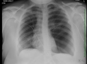
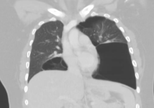
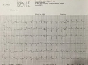
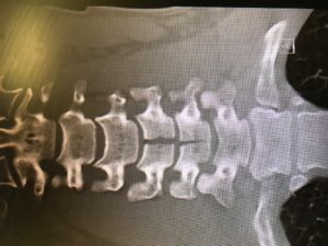
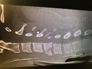
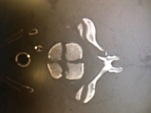
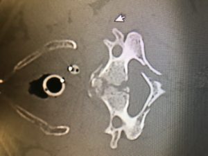
As concern for bronchospasm, the patient was given IM epinephrine and started on a hour long albuterol while history was being obtained.  Bedside ultrasound demonstrated grossly normal myocardial contractility without effusion, normal lung sliding, and a collapsible IVC.  Chest x-ray was unrevealing. After lack of improvement with initial intervention, second dose of epinephrine and fluid bolus begun.  Intial VBG demonstrated hypercapnea and patient was started on BiPAP.  Within minutes, patient showed improvement in respiratory status and appeared more comfortable.  Initial CMP, CBC, CRP, and ESR were unrevealing.   Lumbar puncture was performed after head CT revealed only an arachnoid cyst.  CSF studies demonstrated mildly elevated segs, mildly elevated glucose, negative gram stain and normal protein.  Foley catheter was placed after patient complained of bladder fullness (confirmed by bladder scan) with inability to void.  Neurology and Neurosurgery were consulted from the emergency department, who requested urgent MRI on admission.  Patient was admitted to PICU with ED diagnoses of acute flaccid paralysis and acute neuromuscular respiratory failure.   After admission, MRI was obtained and demonstrated (drum roll please) ischemia vs. infarct from approximately C2-T5 with predominance in the anterior horns.


The literature review of non-traumatic spinal cord infarction is as rare as the pathology itself in the pediatric population and predominately consists of case studies.  Causes typically include but are not limited to hypotension, vascular injury, thrombus, embolus or compression.  Considering many of these etiologies are already scarce in the pediatric population, finding cause proves to be a difficult task.   Pain in neck, back or legs, weakness, tingling, and numbness are commonly described symptoms in pediatric case studies and were present in the patient that presented to us.   Prognosis studies have been performed on primarily adult patients; one such study demonstrated >40% recovery in ambulation amongst the 37 patients that left the hospital wheelchair bound through aggressive physical therapy and rehabilitation programs.[1]   Poor prognostic factors include female sex, advanced age, severity of symptoms, and lack of improvement within 24 hours after infarction.[2]   It appears as though outcomes are primarily dependent on access to physical therapy and supportive care including psychological treatment.   Workup and treatment recommendations if suspicious of atraumatic spinal cord infarct include obtaining an MRI for definitive diagnosis, followed by autoimmune, hypercoagulable, and infectious evaluations. Treatment in the emergency department is mostly supportive and includes addressing any contributory conditions and the management of respiratory concerns.[3]


On follow up, I found that the patient required intubation after exhibiting respiratory decompensation after the MRI was obtained.  Initial treatment with high-dose steroids and IVIG were discontinued when infectious and immunologic work-ups were unrevealing.  The hospital course was complicated by neurogenic bowel and bladder, neuropathic pain, anxiety, intermittent autonomic instability with hypotension, pneumonia and a urinary tract infection.  The patient required tracheostomy and percutaneous gastric tube placement during his hospital stay.  The patient continued to exhibit flaccid paralysis of all four extremities and ventilator dependence on discharge to acute rehabilitation.


Per my own investigation, I was relieved to find that Frazier has both success managing these cases and many unique resources that may positively impact this patient’s course (if you are curious here is some video evidence https://www.youtube.com/watch?v=8o_w174rI3s).  Needless to say, my differential for motor weakness, particularly in pediatrics, will forever be haunted by this case.


[1] Robertson CE, Brown RD Jr, Wijdicks EF, et al. Recovery after spinal cord infarcts: long-term outcome in 115 patients.  Neurology. 2012; 78: 114–121.

[2]Cheshire WP, Santos CC, Massey EW, Howard JF Jr .  Spinal cord infarction: etiology and outcome.  Neurology. 1996;47(2):321.

[3] Spencer, Sandra P. MD; Brock, Timothy D. MD; Matthews, Rebecca R. MD; Stevens, Wendy K. MD.  Three Unique Presentations of Atraumatic Spinal Cord Infarction in the Pediatric Emergency Department Pediatric Emergency Care. 30(5):354-357, May 2014.

Snakes and ladders

Last month I had an interesting Room9 for visual purposes. The story per EMS was jumbled, as it can be from time to time. All we knew was that the patient was a middle aged man who either fell off a roof or jumped off a ladder into a foot and half of water. …agreed. Details were otherwise unavailable. We manage repercussions of injuries, not the causes.

The patent came in intubated, wet, hemodynamically stable. The patient had ketamine en route but was still active with GCS 6T. He required more than your typical sedation to be amenable for the CT scanner. The physical exam showed no motor activity in lower extremities, including to painful stimuli. We noted no step-offs or abrasions. The patient had no signs of trauma other than the motor weakness.He moved his upper extremities and needed restraints due to lack of response to sedation. CT images are below. Most of these images mirror almost exact images from Dr. Ferguson’s lecture on spine fractures, thus I thought it would be good to go over.

As you can see the patient had significant fractures of his cervical spine. Talking with Neurosurgery, the burst fracture is more common in lower thoracic and upper lumbar spine and only is seen in cervical spines to this degree under severe axial load injuries, such as going head first from a significant height (especially when the posterior column has a vertical fracture as seen above).

I’ve always wondered the significance of doing spinal check during the physical exam prior to CT, when they are already getting “manned”. I often feel that if there is real pathology (ie. unstable fractures), won’t palpating (and deep palpation on obese patients) worsen the fracture and theoretically cause neurological issues. I haven’t found much to substantiate that, but it seems to make intuitive sense; I am open to any other opinions / suggestions.

Does anyone want to comment on the type of fractures noted, stable vs unstable (refer to Ferguson’s lecture)?

Are teardrop fractures stable?

Is there any significant retropulsion?

Could you hypothesize flexion vs extension injury?


Answer: Unstable teardrop fracture as well as a burst fracture, and borderline chance fracture (not typical for this cervical spine location).


For further spine related information please look at the links below or Dr. Ferguson’s spine lecture. Ferguson’s is a great source covering the importance of stable and unstable fractures, and a great lecture for interns to go over, especially early on in the year.

Here is a good podcast from Scott W. on less traumatic c-spine injuries:

EMCrit 63 – A Pain in the Neck – C-Spine Imaging and Clearance

And some other good sources:

http://www.aafp.org/afp/1999/0115/p331.html

http://www.paems.org/pdfs/online-ce/Evaluation-and-management-of-acute-cervical-spine-trauma.pdf

Don’t Inhale

Had an interesting case recently while I was working at Jewish Downtown. Approximately 50s male presents in acute respiratory distress. Apparently he rented out some apartments and one of the tenants clogged one of the toilets, I didn’t have a chance to ask how.

Anyway, in an attempt to fix the problem he mixed two plumbing agents: Liquid Fire Drain Opener & some other generic drain-o liquid that he planned to use to clear the pipes. This ended up being a bad idea and a chemical reaction occurred releasing a cloud of chlorine gas. The patient inhaled this and began becoming progressively more short of breath.

While en route, EMS initially had him on a nasal cannula but transitioned him to a non-rebreather as well as a Duo-neb. At the time of his arrival to room 1 the patient was in obvious respiratory distress, diaphoretic, and anxious pulling at the non-rebreather and leads. His O2 sat was from 86%-88% on the non-rebreather with decreased breath sounds diffusely. We elected to intubate the patient with etomidate as our induction agent and rocuronium as our paralytic. After the patient was placed on the ventilator we started the patient on nebulized sodium bicarbonate.

This was something I had never done before – so I wanted to discuss chlorine gas inhalational injuries and the role of nebulized bicarb.

Chlorine gas inhalations typically occur in the setting of chemicals for cleaning an indoor pool. Chlorine gas inhalation rarely progresses to respiratory distress requiring intubation.

In 1994 the Journal of Clinical Toxicology did a two year retrospective review of 86 cases of chlorine gas inhalation and none of the patients progressed to respiratory insufficiency requiring ventilator support.  The most common symptoms are cough, chest pain, and shortness of breath and the chest xray is often unrevealing. In the study all 86 cases received nebulized sodium bicarbonate and none of the patients clinically deteriorated or acutely worsened as a result of the treatment.

While the nebulized bicarb does not reverse or work to eliminate the inhaled chlorine, it helps symptomatically with the cough and SOA. I could not find much information on the MOA or science behind the use of nebulized sodium bicarbonate but it appears to be pretty safe.

The dosing that I found is 4mL of 4% nebulized NaHCO3, and be prepared to explain this to the respiratory therapist because this is not something that they do often. Also this has to come up from pharmacy as code cart sodium bicarb is typically 7.5% so the concentrations are different.

So our patient received his nebulized bicarb once it came up from pharmacy. He was admitted to the ICU and I have not had a chance to follow up on his outcome yet. I just thought this was an interesting case and something that I had not yet encountered and figured I would share for your reading pleasures.

Always keep your differential broad

I had a case in our department that I won’t forget for a while, and it reminded me to keep my differential broad even if the suspected diagnosis seems blatantly obvious.


An early 40’s female presented to our ER about 5 days after an MVC in which she was the restrained driver, where the car rolled onto its side going about 40s-50s MPH. + LOC, + airbags. Paramedics arrived on scene after a while when she was up and walking, and she refused to be taken to the ER. Over the following 5 days, she had near constant neck pain as well as a worsening headache and worsening abdominal and “rib” pain on the lower left side.

She presented to our ER in a hallway bed, where her initial HR was in the mid 80s, but BP was 80s/40s on multiple checks. O2 sat and temperature were normal. Mental status was normal, and there were no physical signs of trauma on her body. She had tenderness to the L lower and lateral ribs, as well as LUQ/LMQ abdominal tenderness, and lower midline C-spine tenderness. I quickly had her placed in a cervical collar, and brought the ultrasound to bedside a performed a FAST, which was negative (to my surprise).

I ordered fluid boluses, trauma labs, type and crossmatch, and planned to send her for a man scan, but her kidney function showed an AKI and therefore had to wait for one fluid bolus before going to the scanner. BP slowly started to trend upwards, not reaching over mid 90s systolic before she went to the scanner. Of note, she did have a slightly elevated white count in the mid-to-upper teens.

My differential? Trauma, trauma, trauma. She has to be bleeding somewhere, she may have a fractured C-spine, intracranial injury, intraabdominal injury, likely splenic laceration. My FAST just must not have picked it up. Given the history and clinical circumstance, I don’t think I was completely wrong for not having anything else on my differential for this hypotensive patient with concerning physical exam findings 5 days out from a serious car accident.

Once her man scan was done, I looked though the scans and noticed her right kidney was heterogenous with contrast enhancement with stranding around it. No fluid in her pelvis, and the rest of the man scan was entirely negative. Radiology soon called and said she had the “worst case of pyelonephritis I think I’ve ever seen”. A urine sample was finally collected after the scan resulted, which was, no longer to my surprise, infected. Upon talking to the patient, she denied any dysuria or frequency, but said her urine was “green” this morning. She never had any suprapubic pain.

That is the story of how I admitted a patient to medicine for pyelonephritis after getting a man scan and diagnosing it on CT. I don’t think I’ll be changing the top item on my differential, but I think I will keep other causes of hypotension and shock on my differential until they are ruled out in cases of delayed trauma presentation, such as this one.

ED Thoracotomy

Link

Resuscitative Thoracotomy

OVERVIEW

  • resuscitative thoracotomy is a thoracotomy performed prehospital, in the emergency department or elsewhere that is an integral part of the initial resuscitation of a patient
  • an alternate term is emergency thoractomy
  • survival 4-33%
  • determinants of survival include mechanism of injury, the location of injury and the presence or absence of vital signs
  • best outcomes in:

-> penetrating chest
-> those exsaunginating from chest tube
-> isolated chest trauma
-> cardiac injuries
-> abdominal trauma that benefits from aortic clamping
-> time since loss of vitals

REQUIREMENTS

  • ETT
  • shock or arrest with a suspected correctable intrathoracic lesion
  • specific diagnosis (cardiac tamponade, penetrating cardiac lesion or aortic injury)
  • evidence of ongoing thoracic haemorrhage

INDICATIONS

Accepted

  • penetrating injury + arrest + previous signs of life
  • blunt injury + arrest + previous signs of life

Relative

  • penetrating injury + no signs of life and CPR < 15min – blunt injury + signs of life in field or during transport -> arrest 15 min
  • blunt injury + no signs of life
  • multiple blunt trauma
  • severe head injury

RESUSCITATION IN TRAUMATIC ARREST

  • 1. Intubate (reverses hypoxia)
  • 2. Insert bilateral chest drains (or thoracostomies)
  • 3. Resuscitative Thoracotomy
  • 4. Limit fluid as this worsens outcome in penetrating thoracic trauma unless haemorrhage controlled
  • 5. Limit inotropes and pressors until circulation restored (will need once defect repaired)

TECHNIQUE

Goals

  • relieve cardiac tamponade
  • perform open cardiac massage
  • occlude aorta to increase blood flow to heart and brain
  • control life threatening thoracic bleeding
  • control bronchovenous air embolism

1. Full aseptic technique*** –> This was recently an issue where the Trauma attending cited both his team and ours in Rm9 for lack of full prep –> masks, surgical gloves, gowns, etc. should be worn when performing this procedure.
2. Scalpel through skin and intercostal muscles to mid axillary line.
3. Insert heavy duty scissors into thoracostomy incisions.
4. Cut through sternum.
5. Lift up (clam shell)

-> relieve tamponade (longitudinal incision through pericardium)
-> repair cardiac wounds (non-absorbable sutures, 3.0)
-> stop massive lung or hilar bleeding with finger (partial or intermittent occlusion may be performed to avoid right heart failure)
-> identify aortic injuries (repair with 3.0 non-absorbable sutures or use finger)
-> consider aortic cross clamping at level of diaphragm (limits spinal cord ischemia)

The Awful Facet of Compare and Contrast Essay Subjects

The Something to complete for Compare and Contrast Essay Matters

Serotonin in small stages reasons a achieve in melancholy. If you’ve got obtained a few products to check or distinction, learn how these are identical and just how these are many different. A new critical difference could be the alternative of conversation treatments.

Compare and Contrast Essay Subject areas Aid!

How to jot down an Essay. Give pupils some assistance on how most useful to determine what is essential. College students are asked for to elucidate, remark on, or evaluate a matter of research inside condition of the essay.

Introduction along with the powerful hook and thesis assertion stays exactly the same. Another eight very simple tips will immediate you thru the method for creating an efficient compare-and-contrast essay that has a specific thing important to mention. essay topics for compare and contrast
It is actually popular to get assigned to compose essays in nearly every discipline of research, not simply when using a composition system.

The Do’s and Don’ts of Compare and Contrast Essay Topics

You’ve acquired a handbook and obtain tips about how to proceed using your composition. To start with, you’ll need to consider one or two compare and contrast essay matters and prefer the ideal just one for you personally. An personal essay can get started which has a thesis, or it could begin with a topic.

Second sizeable component creating an excellent essay is an appropriate textual content construction. The thesis assertion stipulates the key position of or concepts inside of your essay. When he has the knowledge that should enter the essay, she or he ought to just begin the process of producing!Our author can pay with the subjects furnished by your trainer or present other superior options from the occasion you do not possess a various subject. Choosing a subject for Comparison Essay in order to compose a good essay, very first you need to get a superb matter for it, i.e. a subject which helps you to reveal your producing qualities and locate a superior quality quickly. Share an essay on any topic of the collection.

How to jot down a brilliant Compare and Contrast Essay. It is a good deal simpler to compose a comparison essay a few subject matter you recognize a complete whole lot about and also have devoted an abundance of time pondering about. compare and contrast essay topics for 6th grade
Picking out topic options could in fact be tough as you will find a complete large amount of details which often can be in comparison. Nicely, the especially first of all issue that you have to bear in mind is you determine a subject which satisfies you. How to define the suitable Oil On your Automobile. Any high-quality introduction isn’t any in excess of a perfect start off. You do not have to fret regarding your unique particulars which could be considered, as we cope with the issue over a protected community. Do not ever hurry, and work with the define a particular section for the very same time if you ever will require to. Why you should Craft a Reserve. You want to to synthesize your thesis considering the facts in the system paragraphs. You can find no explicit restrict over the degree of text must be composed inside of the essay Crafting Aspect, however it is not mandated to write down over 350 words and phrases considering the fact that examiners customarily dedicate the exact same limitation of your time on nearly every exam get the job done. Now the thesis assertion could possibly be created, and therefore the unique points can become the paragraphs.

You may very well ought to go through up the many journals and circumstance documents to get a superb place. At the exact same time, it might furthermore help you to have a very significantly better image of particularly what exactly are the arguments you would created and regardless of whether they’re related. There are 2 strategies to compose a paragraph, number one, you will find the purpose by stage paragraph and there is the blocked paragraph.It’s not robust to eliminate sight for the notice. Needless to say, you’ll find it likely to demand a nice offer of your time. A lot of people in the present day trust the best will mean of understanding about everyday life is by hearing the advice of relations and acquaintances.

The Another thing to perform for Compare and Contrast Essay Matters

College serves as a time for college students to focus on lessons which might be exclusive towards student’s livelihood goals. Learners will learn about some marvelous details and concepts important for the maturation of the remarkable faculty or school comparative essay. They may sometimes learn the framework with just a quick amount of instruction.

The Concealed Reality on Compare and Contrast Essay Matters

Apparently, you have got to understand gurus from your unique subject if you’d like to write down about personalities. Absolutely the most efficient writers generate each working day, for the exceedingly minimum a tad. College students would not ought to be nervous about bullying any longer.

Trach Management Algorithms/Videos

As a follow up to Dr. Dennison’s presentation yesterday: see this link if you’d like to watch a couple videos on replacement/troubleshooting. They go through passing a new Trach over an airway exchange catheter as well as a Bougie.

http://www.tracheostomy.org.uk/Templates/Videos.html

This link is the algorithms for both Tracheostomy and Laryngectomy: http://www.tracheostomy.org.uk/Templates/Algorithms.html

Facial Edema

This was an interesting presentation from a Peds shift.

15 y/o AAM with no significant medical history who presents with facial swelling. Patient noticed significant swelling to the left side of his face upon awakening in the morning. The swelling involved his entire left cheek, inferior lid of his left eye, upper lip and part of his right cheek. The patient denies any pain, tongue swelling, voice change, difficulty breathing or swallowing, fevers, recent ill symptoms (cough, congestion, vomiting, diarrhea), dysuria, hematuria, rectal bleeding, sore throat, ear complaints. He denies any new exposures including new medications, new soaps, detergents, animal exposures, environmental exposures, recent travel, insect bites.

PMH:none. PShx: had 4 wisdom teeth removal 1 month prior (finished antibiotics), no other recent surgeries or dental work. No EtOH, drugs. No current medications. No known allergies.

Vitals: 97.8, 90, 110/70, 18, 99 % on RA

Exam: HEENT- moderate swelling of the left buccal area, inferior lid of the left eye, upper lip. Mild swelling to the right buccal area. No erythema or palpable areas of fluctuance. No swelling surrounding the right eye. No conjunctival injection. No erythema within the ears, TMs normal. No mastoid tenderness. No lingual swelling, no erythema within the mouth or palpable areas of fluctuance. No signs of infection from previously removed wisdom teeth. No posterior oropharyngeal swelling or uvular deviation. No lymphadenopathy.

Heart- normal. Lungs- clear, no wheezing or stridor. Abdomen- normal. No CVA tenderness.

Treatment started with Benadryl for possible allergic reaction. Basic labs obtained and urine for possible nephrotic syndrome. WBC-17, otherwise normal. Urine with 200 protein, no RBC or WBC- nephrology consulted and recommended repeat POC labs as outpatient and follow-up in clinic, but no intervention at this time. Patient had mild improvement with Benadryl. Discharged home with Benadryl and steroids.

Patient re-presents 6 hours later (just came back for my shift the next day)

Facial swelling has worsened. Now involves bilateral buccal areas, bilateral lower eyelids and upper lip. No fevers, no difficulty breathing, no dysphagia. Patient had taken 1 repeat dose of Benadryl at home and had not started steroids yet. No other changes in HPI except patient mentions some bleeding from the inside of his upper lip. Upon exam, patient has some bleeding and purulent drainage from the gumline of his left central incisor. No palpable fluctuance, but able to express drainage with pressure to upper lip.

Labs obtained: WBC 17, CRP 1.6, ESR 41. UA- minimal protein. All other labs unremarkable. CT face with contrast obtained showing left central incisor periapical abscess with cortical erosion as well as extensive cellulitis of the midface. Also some concern for phlegmon within the paranasal sinus. ENT, OMFS, and finally pediatric dentistry consulted. Patient admitted for IV clindamycin, Unasyn for cellulitis and dentistry consult for possible root canal versus tooth extraction.

Bottom line: Odontogenic infections can cause orofacial infections and rarely but more importantly peripharyngeal space infections as well as jaw osteomyelitis. If concerned about deep facial infection, CT face is warranted. Treatment includes draining of pus from abscesses (either through I&D or needle aspiration) and culture as well as antibiotic therapy. Common regimens include a penicillin plus metronidazole, clindamycin, augmentin, or unasyn depending on disposition. Dentistry should be involved whether through consult or outpatient follow-up for root canal versus tooth extraction.