Good overview of definition, etiology, clinical presentation, diagnosis, and management of kidney stones.
Discussed options for imaging including US and role of non-contrast vs. contrast CT scans.
Most stones pass spontaneously, but prompt recognition of obstruction with infection is critical because delayed decompression can lead to sepsis and permanent renal injury.
Fournier’s Gangrene – Dr. Coleman
Time is tissue! Fournier’s gangrene is a surgical emergency – recognize it early, start broad-spectrum antibiotics, and obtain immediate surgical consultation.
Diagnosis is primarily clinical – do not delay treatment for imaging if suspicion is high.
Usually polymicrobial infection (aerobic and anaerobic bacteria)
Discussed role of clindamycin/linezolid in toxin production inhibition
Urologic Devices – Dr. Shaw
Reviewed case of patient who presented with AMS, electrolyte abnormalities, AKI and infection from post-operative foley catheter placement.
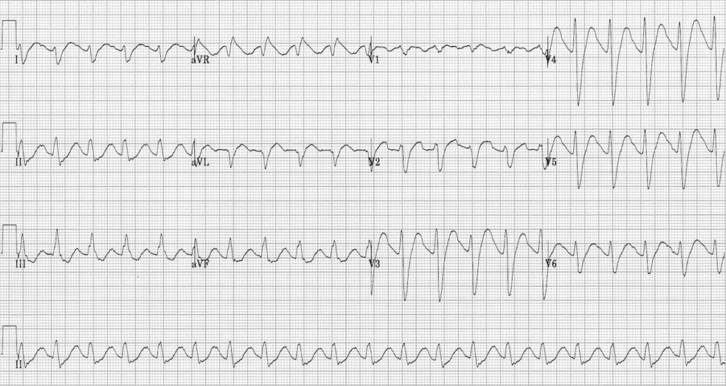
Reviewed EKG findings of hyperkalemia and hyperkalemia protocol – Calcium, Insulin + Dextrose, IVF, Beta-2 agonist
Great review and “show and tell” of multiple urologic devices:
Foley catheter: discussed types, indications (acute urinary retention >300cc PVR, gross hematuria with clots, GU trauma, critical illness/strict I/O), use and troubleshooting
Suprapubic catheter: discussed indications (GU trauma, inability to pass standard foley in setting of retention), placement and exchange (<6 weeks, call urology, >6 weeks, replace)
Takeaway: clinically, many STIs look very similar and do not always present classically; important to recognize and send off testing.
Discussed multiple GU emergencies that may present, specifically with acute urinary retention.
Sepsis Updates – Marianne Dorsey, RN
Reviewed Sepsis Continuum
RN can initiate TIO Sepsis Alert for 2 or more SIRS, suspected or confirmed infection, with or without organ dysfunction Sepsis Alert paged overhead RN to notify MD
Sepsis Time Zero – earliest time that patient meets criteria OR time documented by MD
Reviewed 3 hour and 6 hour sepsis bundles including Tissue Perfusion Reassessment
Reviewed Sepsis PowerPlans (Med Sepsis_ULH)
Document reason for withholding or not giving full 30 mL/kg IVF and excluding criteria for not ordering sepsis bundle
Medical Director Update – Dr. Ross
Discussed new NES study on BP goals in hemorrhagic strokes
Discussed a case on hyperkalemia and reviewed the hyperkalemia order set
Discussed goals for stroke patients including stroke team pre-notification if concern for LVO PTA and TNK feedback
No more fast-gut sutures, plan to use vicryl rapide instead
Alert charge nurse about any concern for Decon – pesticides, acids/bases, toxic industrial materials, gasoline
Methemoglobin refers to ferric form of hemoglobin (Fe3+) that is unable to bind O2
Suspect in any patient with cyanosis not responsive to supplemental O2
Causes:
Rarely congenital, NADH reductase deficiency – unable to reduce ferric to ferrous iron (Fe3+ → Fe2+)
Exposure to oxidizing agent
Benzocaine and other local/topical anesthetics
Nitrites, nitrates, well water (nitrates)
Antimalarials
Quinolones
Dapsone
Aniline dyes
Phenazopyridine
Sodium nitrite toxicity
Evaluation:
Oximetry is inaccurate because methemoglobin (MetHb) absorbs light at both 660 nm (red) and 940 nm (infrared) wavelengths approximately equally. This causes the pulse oximeter to fail to distinguish it from oxygenated hemoglobin, resulting in a characteristic, fixed, and falsely low SpO2 reading of around 85%,
Lack of improvement in oxygen saturation with high-flow oxygen plus discrepancy in oxygen saturation >5% between measurements by ABG vs pulse oximetry (“saturation gap”)
Diagnosis confirmed by measuring level of methemoglobin in blood
Normal PaO2
SpO2 will initially be falsely elevated
Once MetHb >30% → SpO2 will fall to 80-85%
Mid-80s SpO2 due to light absorption of both oxyHb and deoxyHb
“Chocolate brown” color of blood
Management:
High-Flow Oxygen
Methylene Blue
Treat if symptomatic OR asymptomatic with MetHb >25%
1-2mg/kg Methylene blue IV over 5min; repeat dose if no effectImprovement seen within 20min
Contraindicated in G6Pd deficiency (may cause hemolytic anemia)
Other treatment modalities
Exchange transfusion for symptomatic methemoglobinemia in patient with G6PD deficiency
Hyperbaric O2 when methylene blue ineffective or contraindicated
IV hydration and bicarbonate for metabolic acidosis
Inflammatory disease occurring 2–4 weeks after an untreated group A strep infection (strep throat or scarlet fever), primarily affecting children aged 5–15
Connective tissue of heart, joints, CNS, subcutaneous tissues are targeted by immune reaction
ASO (Antistreptolysin O) titer Elevated 1week to 1 month after GAS infection
Anti-DNase B titer is elevated longer than ASO and may be useful for patient presenting later than 2-4 months or with a negative ASO
ECG – Looking for prolonged PR or other AV block
CXR
Echocardiogram
Management:
Eliminate underlying streptococcus bacteria with antibiotics (penicillin), reducing inflammation, pain, and fever with aspirin or other NSAIDs. Consider corticosteroids for heart inflammation.
Long-term, secondary prevention with regular antibiotic injections (Penicillin G IM x1 month) to prevent recurrence and heart damage
Tumor Lysis Syndrome
Overview:
Occurs when large numbers of cancer cells die and release their contents into the circulatory system
Usually with onset of new chemotherapy or high tumor burden.
Air enters pulmonary veins → systemic embolization
Presentation (often within minutes of surfacing)
Neurologic symptoms:
Stroke-like deficits
Seizures
Altered mental status
Paralysis
Vision changes
Other symptoms:
Chest pain
Dyspnea
Cardiac arrest
Treatment:
Same as severe DCS:
100% Oxygen
Hyperbaric recompression (urgent)
IV fluids
3. Barotrauma
Occurs when air spaces cannot equalize pressure.
Ear Barotrauma (Most Common)
-Severe ear pain with descent. If ears are not equalized, pressure builds and eustachian tube collapses. Clear ears on water entry and often during dive.
Symptoms:
Ear pain
Hearing loss
Vertigo
TM rupture
Exam:
Hemotympanum
TM perforation
Treatment:
Analgesia
ENT follow-up
Avoid further diving
Give antibiotics only for perforated TM
Sinus Barotrauma
Symptoms:
Facial pain
Epistaxis
Sinus pressure
Treatment:
Decongestants
Analgesia
Pulmonary Barotrauma
Causes:
Pneumothorax
Pneumomediastinum
Arterial gas embolism
Symptoms:
Chest pain
Dyspnea
Subcutaneous emphysema
Treatment:
Manage pneumothorax
Oxygen
Hyperbaric if embolism suspected
4. Nitrogen Narcosis
Occurs at depth >30 meters (~100 ft).
Symptoms:
Euphoria
Poor judgment
Confusion
Impaired coordination
Treatment:
Ascend to shallower depth
Symptoms resolve quickly, it is safe to continue diving after resolution of symptoms.
Immediate ED Management
High-flow oxygen
IV fluids
Neurologic exam
Call hyperbaric center
Transport if needed
In the U.S., consultation is often through:
Divers Alert Network, they assist with hyperbaric referral and organizing transfer
Board Pearls
Any neurologic symptom after diving = assume AGE or DCS → hyperbaric therapy
Symptoms within minutes of surfacing → think AGE
Symptoms hours later → think DCS
Joint pain after diving = DCS Type I
Stroke-like symptoms after diving = AGE until proven otherwise
Immunosuppression and Transplant
Common ED patients:
Solid organ transplant
Bone marrow transplant
Chemotherapy
Chronic steroids (>20 mg prednisone daily for >2 weeks)
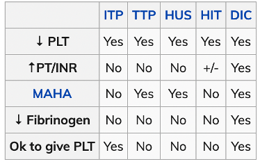
Insufficient ADAMTS-13 activity allows vWF multimers to accumulate in microcirculation which leads to platelet aggregation/thrombocytopenia and hemolysis of RBCs.
Risk Factors:
Congenitally deficient ADAMTS-13 activity AND:
Pregnancy OR
Infection OR
Inflammation OR
Medication use (quinolones, ticlopidine, clopidogrel)
Testing not always needed, UTD recommends CBC, CMP, ESR/CRP, C4 level (c4 level +/- c1 inhibitor antigen testing used for future diagnosis, not part of ED management)
Imaging not typically needed unless concern for concurrent dx (i.e. infection, abscess)
Anticipate difficult airway, have backup available if possible
Be prepared for being unable to oxygenate and ventilate, be ready to perform cric
Be as gentle as possible- tissue is friable, irritation will cause more swelling.
Consider intubation early-angioedema can surprise you and progress very rapidly.
Consider intubation very early in post-TNKase cases, as cric could be devastating
Digital (tactile) intubation strategy: Use the non-dominant index/middle fingers to palpate the epiglottis, directing a boujie or ETT into the trachea.
Dispo:
No consensus on timing needed for obs. If stable need to be observed until signs of improvement. Most non-allergic angioedema does not fluctuate in severity.
Consider admission: If partly allergic picture, high risk body part (airway), poor social situation
Consider transfer: Low resource facilities, no advanced airway clinician except you, consider airway compromise en route (sometimes early intubation is the safest option prior to transfer)
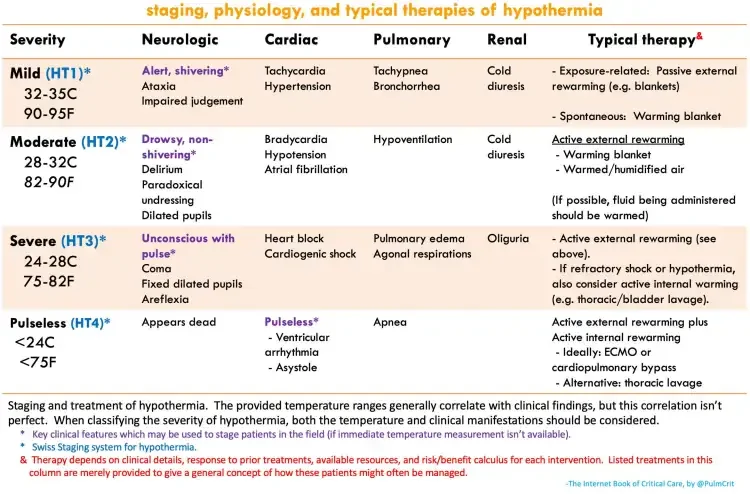
Modifications to standard ACLS algorithms in hypothermia
Medications may fail to be metabolized and accumulate. Therefore, avoid repeated doses of medications.
Defibrillation may be unsuccessful until the patient is rewarmed. Thus, serial shocks for VT/VF arrest are unlikely to add benefit. As the patient rewarms, further attempts may be made at defibrillation.
Rewarming patients with frostbite:
Goal for rapid rewarming. 40-42C is around 104-106F or about the temperature of a hot tub. This will rapidly rewarm but will not cause burns. Anything less will not rewarm tissue sufficiently. It is important NOT to rub/massage the affected digits, as the crystallized tissue is fragile and this will cause further cellular damage.
Tx: rewarm, liberal pain control, dressing/wounds care, reassess
High altitude cerebral edema (HACE) = always descend!
DERM
Rheumatic fever = Jones criteria = erythema marginatum (weeks later)
Core competencies
Interpersonal and communication skills: capacity
not competence, an informed choice, alcohol use
Level A recommendation: established evidence, multiple RCT, meta-analysis
Level B recommendation: some evidence: single RCT; multiple population
Most specific finding is a ruptured aneurysm with retroperitoneal hematoma
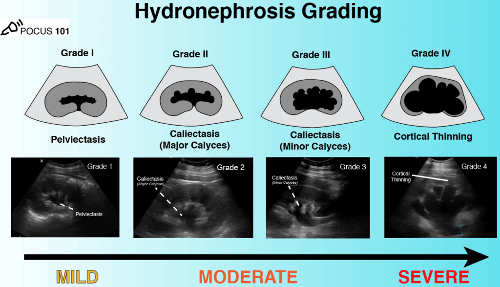
Hydroneprhosis
Cholecystitis
Measure CBD inner wall to inner wall; normal <5mm
Retinal detachment is tethered to the optic nerve
Vitreous detachment is mobile and can lead to retinal detachment
(+) HCG
Discriminatory zone 1500 IU/L
IUP confirms requires intrauterine gestational sac plus yolk sac
gestational sac alone = pregnancy of unknown anatomic location
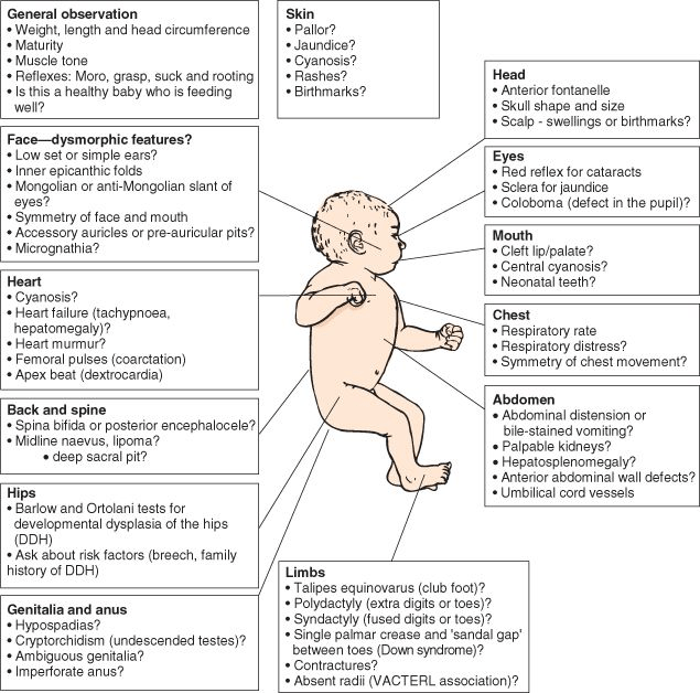
The newborn exa
examine naked and head to toe, every time (think mini trauma assessment with more steps)
Newborn well-care
The nursery
If there is suspicion you may need to tap the baby, you MUST ask about vitamin k administration
should receive hep b, erythromycin ointment to eyes, and vit k IM within 1-2 hours of birth
Feeding
newborns should take a minimum 1-2 oz of formula or breastmilk every 2-3 hous (including overnight). Normal 19 kcal/oz formula should be mixed 1 scoop of formula to 2oz of water. Water first then formula.
Administer IV sodium bicarbonate until you see changes in EKG
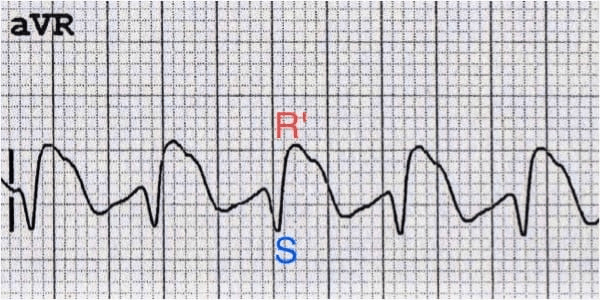
1-2 mEq/kg as intravenous push every 5 minutes as needed for termination of wide-complex tachydysrhythmia or prolonged QRS interval >120 milliseconds (ms).
Management- fluid replacement, temperature control, +/- steroids
Dispo- trauma/derm/burn unit
Dr. Firquin- Pediatric respiratory emergencies
Croup- URI symptoms, fevers, stridor, barking cough, symptoms worse at night, hypoxia is uncommon
Corticosteroids (0.6mg/kg with max 16mg), racemic epinephrine
Bronchiolitis- URI symptoms, tachypnea, peak at <12months, retractions, poor PO
HFNC, suction
Foreign body aspiration- cough, stridor, tachypnea, sudden episodes
XR, bronchoscopy
Asthma- wheezing, cough, triggered by viral illness
Albuterol, ipratropium, steroids
Magnesium- side effects of bradycardia and hypotension
Epinephrine IM 0.1mg/kg for severe cases
Terbutaline for severe cases
Consider CXR on first time wheezer
Dr. Smith- Cutaneous ulcers
Diabetic foot wound- neuropathic ulcer over bony prominences of foot, cover for MRSA and pseudomonas for infection, non-infected wounds can be discharged with outpatient follow-up
Venous stasis ulcer- caused by poor venous return, shallow ulcers with irregular borders, wet to dry dressings, elevation of leg, compression stockings, oral antihistamines for itching
Pressure ulcer
Stage 1/2- dressing changes, padding for pressure relief
Stage 3/4- debridement, wound care, ongoing pressure relief
Arterial insufficiency ulcer- usually able to be managed as outpatient as long as patient has good capillary refill, pulse
Pulseless- CTA, trauma consult
Calciphylaxis- ESRD patients, deposition into arteries, rapidly progressive lesions
treatment with calcium control and possible surgical intervention
The main route for toxicity is through ingestion. Onset of symptoms can occur as early as 15 minutes after ingestion ranging from sweating and agitation to seizures, cardiac arrhythmias, and cardiopulmonary arrest [1,2].
Benzos, phenobarbital for seizures in camphor
Amitriptyline
Na channel blockade, CNS, Cardiac, anticholinergic
Long QRS
Na bicarb tx for cardiac dysrhythmias
Benzos for seizure
Lomotil
Imodium
Antimuscarinic and opioid symptoms
Tx naloxone maybe gtt
Need admission for 24hrs
Ethylene glycol
Antifreeze degreaser engine coolants
Oxalate binds with calcium and deposits causing hypocalcemia
Thiamine, pyridoxine,
Tx: fomepizole, dialysis
Dr. Platt – Syncope
Reflex, orthostatic, cardiac
Initial eval – detailed history: prodrome, extertional, position, family history, chest pain, palpitations, hypotension at triage
Physical exam (cardiac, neuro, orthostatics
EKG
High Risk: syncope during exertion or while supine, abnormal cardiac exam, family history or sudden cardiac death, short/ absent prodrome
RBBw/ LAFB syncope no prodrome and exertion needs admission.
Risk stratification tools
San francisco syncope rule: CHF, hct, ecg abnormal, sob, SPO2
CHESS risk factors
Predicts 30 day serious outcome (arrhythmia death, cardiac event)
Scoring history, ekg findings, troponin, ED diagnosis
![Room9er ["Room Niner"]:](https://room9er.com/wp-content/uploads/2020/03/cropped-Screen-Shot-2020-03-08-at-3.16.16-PM.png)