Public health for adrenaline junkies

Public health is associated with the least interesting courses of medical school (epidemiology and biostatistics). It has been linked to ED frequent flyers and psych hold patients (social and behavioral health). One might say it’s an integral part of vague clinical decision rules and flip-flopping society guidelines and Press-Ganey scores.

It’s also something us ED docs do every day, whether we want to or not. There’s no reason why we shouldn’t try to understand it and do it better.

Kiran and I have been developing a website dedicated to public health for ED personnel. It’s a FOAMed site. Our plan is to get it tied in with sites like EMCrit, ALiEM, LITFL, and the like. It’s still a very young project, but there’s a couple articles posted for you to glance at.

If anyone is interested in population health, there’s a ton of uncharted territory and we’d welcome the collaboration. Just let us know how you want to be involved. Also, tell your friends.


Burnout

Brief description of burnout types. Not medically specific. Follow the links as well.

1. Overload: The frenetic employee who works toward success until exhaustion, is most closely related to emotional venting. These individuals might try to cope with their stress by complaining about the organizational hierarchy at work, feeling as though it imposes limits on their goals and ambitions. That coping strategy, unsurprisingly, seems to lead to a stress overload and a tendency to throw in the towel.

2. Lack of Development: Most closely associated with an avoidance coping strategy. These under-challenged workers tend to manage stress by distancing themselves from work, a strategy that leads to depersonalization and cynicism — a harbinger for burning out and packing up shop.

3. Neglect: Seems to stem from a coping strategy based on giving up in the face of stress. Even though these individuals want to achieve a certain goal, they lack the motivation to plow through barriers to get to it.

In a roundup of 11 ways to beat burnout, 99u offers this breakdown of the three main types of burnout, per the Association for Psychological Science.

Pair with this essential read on how to transcend the “OK plateau” of work, then see how sleep factors into the equation.

Here is the article itself from APS.


Reference:

http://explore.noodle.org/post/83726976259/1-overload-the-frenetic-employee-who-works?utm_content=bufferef345&utm_medium=social&utm_source=twitter.com&utm_campaign=buffer

The Hypercoagulable Liver Failure Patient?

Hey Guys,
Just listened to a portion of April EM:RAP (so if you’ve heard it already here’s a little repetition) and was surprised to hear their Notes from the Community Section about coagulopathy in Liver Disease, which basically informed me that many liver patients are at just a high of risk of thrombosis as they are of bleeding.

3 Articles were referenced:
– Tripodi A, Mannucci PM. The coagulopathy of chronic
liver disease. N Engl J Med. 2011 Jul 14;365(2):147-56.
– Villanueva C et al. Transfusion strategies for acute upper gastrointestinal bleeding. N Engl J Med. 2013 Jan 3;368(1):11-21.
– DeLoughery TG et al. Invasive line placement in critically ill patients: do hemostatic defects matter? Transfusion. 1996 Sep;36(9):827-31.

Summary:
– The liver makes both procoagulant and anti-coagulant proteins which can be actually reduced close to equally rendering the patient basically in equilibrium (thus not so hypercoagulable).
– No study has shown that coagulation defects predict issues with procedures (but the experience of the clinician performing the procedure does).
– INR is not standardized in Liver Failure patients (like it is with Warfarin) and thus is not that helpful.
– PT & PTT may also not be helpful in patients with liver failure (due to the variability of loss of clotting factors)
**Fibrinogen may be low in liver failure patients, and you may consider replacement of this with Cryoprecipitate (10 units of Cryo increases the Fibrinogen by 100mg/dl).
– Raising intravascular volume with pRBCs may make them bleed more.

For the full references & discussion see the EM:RAP written summary. Perhaps this is something we should discuss with our GI colleagues and/or MICU people. Any thoughts?

Wellens’ Or Not?

A middle aged male presented for evaluation of AMS; he had agitation, confusion, and tremors. He has a history of Bipolar disease and schizophrenia as well as HTN.  Vital signs were all stable on presentation and within normal limits.  On exam he was oriented only to name, but not place or time. Neurological exam was normal, with the exception of tremors.  Med list includes Haldol Injection, Lithium, Benztropine, Olanzapine, and Propranolol.  At this point, I was not quite sure what is going on with him, so I had a bit of a shotgun approach.  Initial EKG revealed the EKG below.

20140325_223727

It appears to be similar to Wellens’ syndrome but not consistent with my gentlemen’s symptoms.

A quick literature search revealed a case report showing lithium induced EKG changes, similar to his EKG above.  Further supporting his lithium induced changes in this scenario is no clinical findings to suggest ACS, and a negative troponin 3x.  He was ultimately admitted and treated for lithium toxicity, without any cardiac complications.

EKG changes seen in lithium toxicity:
– ST elevations (1 other single case report)
– QT prolongation
– non-specific ST segment changes/T-wave abnormalities

FOAMed and Social Media in Education

At the CORD Academic Assembly there was a lot of talk about FOAMed and Social Media. I am on the social media committee with CORD and we have some informative projects on the horizon.

Here is a blog post detailing some of the FOAMed workshop with some How-To information.

Then Joe Lex and Nicholas Genes posted PRO and CON articles on the question “Is FOAMed essential to Emergency Medicine education.”

Not long articles, good to be at the forefront of these changes. And good discussion on the pluses and minuses of FOAMed

March EM:RAP Summary

EMRAP_2014_03_March_3.1.14[1]

Hey Guys,
Just some things I learned on EM:RAP this month, and since I’m on admin thought I would post a few things.

The DRE (not the rapper Dr. Dre): from the Fingers & Foley’s section on EM:RAP; reviewed Esposito TJ et al. Reasons to omit digital rectal exam in trauma patients: no fingers, no rectum, no useful additional information. J Trauma. 2005. They had 512 trauma patients at a Level I Trauma Center. “the negative predictive value of both the rectal exam and other clinical indicators was very high: 99%. However if the other clinical indicators missed the injury, so did the rectal exam. It didn’t add any information”
Basically showed added value of a rectal exam to be very minimal, and that ROUTINE RECTAL EXAM IS NOT RECOMMENDED. BTW, this is in the journal of trauma which is well-regarded amongst our surgery friends.

How to identify if patient’s contact lens is still in the eye (e.g. if patient has eye pain and feels like its still in there and can’t find it): Use Fluorescein much like you would for corneal abrasion as it will stain the contact lens as well, allowing you to find it (then you can evaluate for corneal abrasion at the same time). *keep in mind contact lens will be ruined*

Subarachnoid Hemorrhage: from an article in JAMA in Sept 2013 with 10 university affiliated Canadian EDs. Tried to come up with a decision rule for SAH and basically came up with:
The decision rule including any of age 40 years or older, neck pain or stiffness, witnessed loss of consciousness, or onset during exertion had 98.5% (95% CI, 94.6%-99.6%) sensitivity and 27.5% (95% CI, 25.6%-29.5%) specificity for SAH. Adding “thunderclap headache” (ie, instantly peaking pain) and “limited neck flexion on examination” resulted in the Ottawa SAH Rule, with 100% (95% CI, 97.2%-100.0%) sensitivity and 15.3% (95% CI, 13.8%-16.9%) specificity.
**Key points: this decision rule is more to identify high risk patients and the high risk symptoms. Sensitivity was very high, but specificity very low. Also keep in mind there are other important causes of headache to keep in mind**

Biphasic Reactions in Anaphylaxis: from Annals of Emergency Medicine in November 2013. A chart review was performed over 5 years and had ~500 pts with anaphylaxis, but also documented allergic reactions. Found biphasic reaction was extremely rare (2 cases while in the ED, and 3 out of the ED), with a rate of 0.4% while in the ED. 6% bouncebacks in the anaphylaxis group, none of which died and none of which came back in anaphylaxis. This study was limited in that it was retrospective, and there was variability in the outpatient management of these patients (unclear who was DC’d with what if any medications)
Rosen’s states corticosteroids can be helpful in reducing risks of protracted anaphylactic reaction and biphasic anaphylaxis (7-10 day course). H1 & H2 antihistamines are helpful in reducing some of the symptoms of anaphylaxis. 5-Minute Emerg Consult recommends Epi-Pen for those with anaphylaxis.
No Formal observation time has been established, though Rosen’s says 2-6 hours; with longer observation times/admission for those with prolonged reaction or requiring multiple dose epinephrine

Sorry this is so long, but I’m on Admin so thought I’d post something. Attached you’ll find the March Written Summary with all the articles they referenced. Hope this is helpful.

The Trauma-Stroke Eval

Middle age male brought into Room 9 s/p crush injury. The patient was reportedly crushed against a box truck and a wall for a short period, but no prolonged extrication.

He complains of LUQ chest pain, pleuritic in nature, and denies abdominal pain. He was ambulatory after the event. PMH only significant for HTN, otherwise no PSH, no allergies, and takes lisinopril and hydrochlorothiazide.

He is AOx3, moving all extremities, well appearing in Room 9, and acting appropriate. He goes for man scan as he did strike his head and although can give me all the details of the accident he reports LOC.

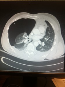
Upon return from the CT scanner his VS are stable, and is placed in a regular ED bed.  Image review revealed multiple rib fractures, and suspicious appearing spleen.  Radiologist confirms grade 1 spleen laceration, but no active extravasation, as well as bilateral rib fractures.

While evaluating another Room 9 patient, the nurse came to update me that the patient was now unresponsive and foaming at the mouth.  I told her to give Narcan, thinking it could be iatrogenic analgesia toxicity, and I would be right there.

I arrive in the room, and he does NOT look right. He is foaming at the mouth, the narcan certainly changed his pupil size when she gave it, but did nothing to his mental status. Sternal rub, nipple pinch, and ammonia capsule all failed to appropriately arouse him.  The trauma resident is now at the bedside as well and just as perplexed as this isn’t what I described on the phone. I call radiology ask them to re-check his CT head but they said no, they see nothing. I again re evaluate him and now he will move only his L side, and follow commands on the L side of his body.

Well…..crap, this is dysarthria R sided hemiplegia. Stroke paged, obtained stat CTA head and neck, and then straight to Room 9 to intubate him. See the CTA imaging below:

CTA

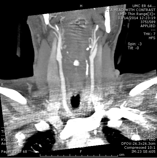
Carotid artery dissection…

He obviously is not a tPA candidate, but he did go for an intervention procedure with the stroke service.  He did have a clot retrieved, but I have visited him on the floor a few days later and he had no improvement.

He went on to develop cerebral edema as well and had a large hemicraniectomy. He also had an ex-lap for an increase in abdominal distension, and found to have pancreatic ascites (3L removed from the abdomen). The spleen did fine.

Looking back, his mechanism did not support a reason for him to dissect a carotid. He had no external signs of injury, he had no neck pain, my guess is he did have a whiplash type injury but again, not something I expected. In addition, this man had a normal neuro exam upon presentation, full strength, no numbness/tingling with the exception of reporting headache, which I thought was from him striking his head.

After a lit review, there are a few teaching points I want to highlight from this case.

Traumatic Dissection (carotid, vertebral, spinal arteries)

– consider it with hangings, significant head/neck trauma, hyper-extension injuries, lateral rotation injuries of the head, base of the skull fx, c-spine fractures(especially those with displacement or involvement of the transverse foramen,vertebral body), lefort fractures (types II and III), or seat-belt sign over the neck
– Sign/Symptoms vary greatly. Stroke like symptoms are concerning but can be as generalized as headache/migraine, neck pain, neck hematoma, blindness, aphasia, weakness/sensory loss, Horner’s syndrome, tinnitus, CN deficits, diplopia, locked in syndrome, ataxia, vertigo, dizziness

Utility of a CXR

Hey Guys,
Thought I’d post this as I feel it was a mistake on my part, though fortunately no harm came from it.

50 year old guy this past Saturday (which was a ridiculous shift full of drunks and unhelmeted mopeds and motorcyclists with some very sick people) who was an unhelmeted moped rider going reportedly 60mph and wrecked while drinking alcohol and somehow managed not to make their way to Rm 9.
BP: 125/73. HR: 86. RR: 18. O2 saturation: 91% on room air. Temp: 97.6 F (oral)
A&Ox3, c/o shortness of breath and diffuse chest tenderness. Not really any obvious bad looking signs of trauma and had been log-rolled prior to my eval (triaged about 1hr 45min before I saw him).

I saw him, in no respiratory distress, maybe some expiratory wheezing on my exam, but with breath sounds on both sides and no crepitus that I could feel to his anterior chest. I finish my eval and just order a MAN scan based on his Hx and due to his drinking alcohol (EtOh 292)
Patient is taken to CT at 23:05 (about 2 hrs after I ordered them) and the nurse grabs me after the scan and tells me I need to look at his chest CT (only his C-spine images were up at the time which showed me all kinds of SubQ emphysema).

Chest CT Pneumo

Just a lesson learned; when it’s super busy like that shift was is when we need to be the most cautious and really think critically about the things we need to do, and not be in a hurry when we’re putting orders in.
The guy is doing well so far (and not intubated) and in THIS case no harm was done, I put a chest tube in without difficulty. But I really should have gotten a CXR to start.
His injuries included Bilateral 1st-6th Rib Fx’s, Pulmonary Contusions, Mediastinal Hematoma, Manubrium Fx, C7 Fx, T4, T7 Fx.