A-fib with RVR + sepsis + hypotension = conundrum

How do we slow the rate without lowering the BP? Will slowing the rate even help the BP? How do we raise the BP without speeding up the rate?

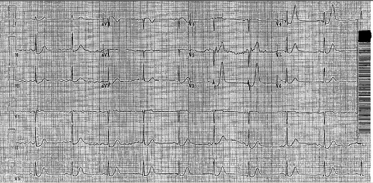
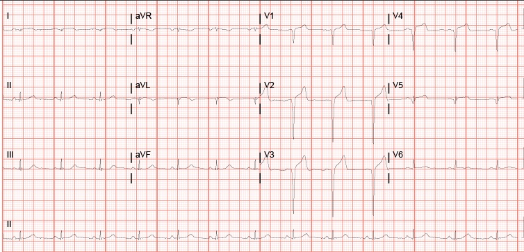
The patient who inspired this post came in for a bowel obstruction. Cards was initially consulted for possible new a-fib, but there really wasn’t much to do from a cardiac standpoint. THEN he perforated his bowel and went for emergency surgery, where he required pressors and went into a-fib with RVR. He was packed open and taken to the SICU, where he was hypotensive to SBP 70s, tachycardic to 160s, and intermittently hypoxic.

If you want to skip my thoughts on the case and head straight for the facts, here is an interesting article about a-fib in critically ill patients. It talks about the various options for management and the pros/cons of each.

If expert opinion is more your style, try this.

Of course, there is also controversy around slowing down a-fib when it is caused by sickness. Should we let the body do its thing to try and compensate? Here’s one article that suggests maybe we shouldn’t get so hung up on rate control in sick people.

Now back to the case. The surgery resident and I were of the mind that slowing the rate and organizing the rhythm should help with cardiac output. There are several reasons why this logic still seems to be in the majority.

– With a-fib you lose atrial kick. That little extra oomph from the atrium may not seem like a big deal, but it can have a significant impact on cardiac output. Here’s a fun article from 1965 that shows a 53% increase in cardiac output from converting a-fib to sinus (using quinidine because 1965). The results have obviously been redemonstrated in more recent studies, but how often do you get to reference quinidine? Not that often.

– Diastolic filling is important for stroke volume. With any tachyarrhythmia, less diastolic filling time means lower stroke volume. However people who do “math” would argue that increasing the rate would likely keep cardiac output about the same. This logic holds up with regular rhythms, but studies show that irregular rhythm decreases cardiac output compared to regular rhythm. Here’s one such study.

– In real life, we went for amiodarone and electrical cardioversion. From the a-fib in critical care article above, it seems like that’s still the best option.
Here’s a good article I wish I had read before embarking on a cluster of a cardioversion. P.S. Put the pads on correctly. Anterior-posterior pads definitely worked better for this 350 pound patient. Anterior pads = fail x3. AP pads = success!

– It’s worth noting that this is not one of the scenarios when you’re worrying about giving someone a stroke with cardioversion. This guy’s risk of death was much greater than his risk of stroke.

Another consideration for this situation was the choice of pressors. Eventually the patient ended up maxed on pretty much all pressors, but that may not always be the case.

Surviving Sepsis guidelines are all about Levophed as a first line pressor, which is usually a great option. But guidelines are just guidelines. How many of our patients are otherwise 100% healthy and just have a little sepsis? Not that many.

– In this case, I think phenylephrine may have been a better first option. Pure alpha agonist activity vasoconstricts without Levophed’s cardiac effects, which probably didn’t do us any favors with the RVR.

– Based on the EMCrit blog above it also seems like phenylephrine might have allowed us to use a beta blocker without worrying about blocking the effects of the pressor or a calcium channel blocker without exacerbating hypotension. Thoughts?

– Anyone have other ideas about pressors in this scenario?

There was a lot more to the case after that, but this post has already ended up way longer than I intended. In the end, it was a 350 pound unhealthy guy with a less than ideal heart, so unfortunately, his family ended up withdrawing care. I doubt anything we could have done would have changed his outcome, but maybe there’s something to learn from it that will help someone else someday. I’ve talked to several people about this case and gotten different opinions from each one, so I figure why not open it up for a few more opinions to really confuse clear things up.

Everything including the lipid sink

80+ yo gentleman with PMH of COPD, CHF, CAD, CKD, initially hospitalized for urosepsis and NSTEMI with a worsening AKI, and baseline CKD. Tuned him up and sent him to the floor where he accidentally received an overdose of his carvedilol. Got the call that the patient was hypotensive to 70s systolic, bradycardic to 30s.

Immediately pacer pads were placed to ward off evil spirits
-Started with 1L crystalloid bolus –> no response and with risk factors and pmhx, didn’t want to fluid overload
-Tried atropine (3-4 doses) –> no response
-Tried glucagon 50mcg/kg loading dose followed by 3mg/hr infusion –> still no luck and exhausted all the glucagon in the pharmacy
Running out of options here. I remember reading an article in Annals about using lipid infusions in beta blocker overdoses and figured it was worth a shot.

Started 20% Intralipid bolus through central line at 1.5ml/kg followed by 0.25ml/kg/min for 30min. Maybe it was the lipid infusion or just the combined effects of everything but by 6am, pt’s BP and HR had improved to normal limits and he began talking again.
It was a nice result but one I couldn’t celebrate that much – within a week, patient decided to go to palliative care and that was the last I heard of him.

PEM Chronicles: Rasburicase

     A case I saw  last month led me to utilizing a drug I’d never heard of before in the ED: Rasburicase.

     The drug: a recombinant urate oxidase enzyme, which converts existing uric acid to allantoin. This is key due to the higher solubility of allantoin in urine. Patients with Tumor Lysis Syndrome are at risk of acute renal failure due to precipitation of uric acid crystals in renal tubules and collecting ducts.

The case: a 12 yo F with no PMHx who was transferred from an urgent care center for multiple tender, enlarged lymph nodes and a WBC count of 98.

As we worked her up for a new presentation of a hematologic malignancy, it became evident she fit into the parameters for TLS. Initial uric acid level – 14.5 along with hyperkalemia, elevated serum LDH, and hyperphosphatemia. When Hem/Onc came on board, once labs were back, the first recommendations were hydration and Rasburicase.

Provided below is the article I found that informed me on the treatment and a few key points picked up when using it.

For kids only – The drug hasn’t been approved in the US for adults.

G6PD deficiency – Yes, from the depths of med school knowledge, this condition is a contraindication to using Rasburicase. The hydrogen peroxide it produces as a byproduct can lead to hemolysis. Ask if there is a family history.

Coordinating with pharmacy – Due to the preparation of rasburicase, it should be administered immediately after it has been prepared. IV access should be obtained well in advance.

Type of IV access – In our case, hem/onc was considering emergent dialysis. It may be prudent to discuss this with your consultants to get a head-start on what they’ll need once going upstairs.

Not at your medical facility, a specialized pediatric hospital, or somewhere with this in stock??? As you transfer, consider:

1.) Aggressive IV Hydration. Easy to start getting this started, especially if transporting to the next hospital will require hours instead of blocks.

2.) Allopurinol. Though it isn’t required when Rasburicase is given, allopurinol can’t decrease what is already present, but it can help prevent the formation of more uric acid.

http://www.ncbi.nlm.nih.gov/pmc/articles/PMC1200736/

The elusive S1Q3T3

So this is a case I thought was interesting that I had in the department back in May. We all know that the most common EKG finding in the setting of PE is sinus tach, however the pimp question that is also asked is the finding of S1Q3T3. While I can easily recite the alphabetic-numeric code S1Q3T3 by heart at the drop of a hat, I had never seen one and honestly thought I never would. I thought finding S1Q3T3 was likely as rare as surviving a ED thoracotomy (OK maybe not that rare).  So on to the case.

39 year old female presents to the ED complaining of SOA and cough for the past two weeks. Cough was productive of green sputum, no fevers, and she does complain of some chest pain which sounds pleuritic in nature. Initial vitals HR 118 BP 111/73 RR 16 O2 97% room air. As I get into her PMH she says she has a history of multiple PEs with an IVC filter placed 1 yr ago because apparently she wasn’t very good at remembering to take her coumadin. She has had a hypercoagulability workup which was negative, no recent travel, no estrogen use.

So at this point with a history of multiple PEs, tachycardia, SOA, and pleuritic chest pain I am thinking I am going to scan this lady. Even though she had a IVC placed a year ago, she is still saying all the right things for PE. So while the CTPE protocol was cooking I got an EKG and there it was, S1Q3T3!

S1Q3T3

Needless to say I was pretty excited and immediately showed the rotating intern next to me who clearly didn’t share my enthusiasm. When I compared this new EKG to a past EKG a month ago she did not have the S1Q3T3.During her admission a month ago, when she had a normal EKG, she had a CTPE showing a chronic PE. This time when her CTPE came back the read was Acute on Chronic Pulmonary Embolism. So a month ago she had a chronic PE with a normal EKG and at this visit she had an EKG with S1Q3T3 and a acute on chronic PE. Out of curiosity I dug through her medical history a little bit more and found that this patient had multiple prior admissions for PE with multiple CTPE protocols and EKGs. What I found was that whenever this patient had a CT read of Chronic PE she had a normal sinus rhythm EKG. However, whenever she had a read of Acute on Chronic PE (which was 4 times!) she had a EKG showing S1Q3T3, dating all the way back to 2012. Yea, apparently this lady has been hanging out with a chronic PE in her distal right main pulmonary artery since 2012 and every once in a while she will throw a new small PE, even with an IVC filter.

So after doing a little bit of thinking and a little bit of reading it made sense. The EKG finding of S1Q3T3 is indicative of right heart strain, in this case resulting from an acute PE. So this patient’s heart has adapted to her chronic PE, however every time she throws a new PE she has an element of right heart strain which can be seen on her EKG as S1Q3T3.

I just thought this was pretty interesting to actually see the physiologic and mechanical adaption and strain this patient’s heart was undergoing being clearly demonstrated on her EKG. Also I learned that S1Q3T3 is not like a q wave after an MI in that it stays on the patient’s EKG, it is a finding that comes and goes depending on the patient’s presentation.

Anyways I thought this was a pretty cool little case and figured I would share. Hope you all enjoyed.

The Kitchen Sink

Fairly early into an overnight shift the radio goes off.  EMS is approximately 6min out with mid 30s male undergoing CPR. Per bystanders, the patient was going running from door to door, banging on doors and yelling for help. Shortly after being taken into police custody he was found to be pulseless. Had been undergoing CPR approx 10 min. King airway in place. No improvement with dextrose and naloxone.

The intern on and myself go to Room 9 to get set up. When EMS arrives about 10 minutes later, CPR is still ongoing and rhythm has been asytole/PEA throughout. The story remains consistent with him going from door to door yelling for help but now there is some variation as to whether he was cuffed and then lost his pulse, was found without a pulse, or if he got into a physical altercation with the neighbor and then lost pulse shortly after arrest. There are no signs of trauma and we elected to not invite additional chaos by calling a Level 1.

CPR is continued. King airway switched out for ETT. Accucheck 250’s. Central venous access and bilateral chest tubes placed with no rush of air or blood. Sats persist at 85% on vent, etco2 is in 60’s. He gets epi q3m. A dose of vasopressin and steroids. Istat has K 5.5. Lactate >20. Received 2-3 amps of bicarb. Received calcium. Upon rhythm checks he was primarily asystole except for 2-3 checks with a narrow pea that would not persist. Each time the ultrasound was placed on his heart there was no cardiac activity. My thought process at this time was that he was either excited delirium that I’m not going to be able to do much about or some kind of ingestion/overdose. We called pharmacy and asked for lipid emulsion. His ETCO2 had remained between 40-60 and we were about 40 minutes out from reported time of arrest. I decided if the lipids were unsuccessful that would be when we would call it.

They arrived about 15 minutes later and were infused. By now his etco2 had drifted down to 10 on the most recent pulse check. No changes occurred and TOD was called.

Reasons not to get into prison fights…

Middle aged male transferred from an OSH, accepted by ENT for a mandible fracture.

The patient is incarcerated, and was involved in an “altercation” with other inmates. The incident occurred around 2PM; but he didn’t report any of his pain to the guards until 10PM.  On arrival at the OSH he had multiple contusions to his face/head, lacerations over his hands, and obvious dental trauma.  The patient was also complaining of chest pain – he stated that another inmate had slammed him in the chest with his knee. Despite his age, the patient has a history of previous MI in 2011, cathed at U of L with no stents placed. Takes a baby aspirin, no other meds and no other PMH.

At this point, the patient is about 10 hours out from the incident. Work-up at the OSH with the following: neg CT head and CXR. CT face with a mandible fracture. Labs notable for WBC 17.8, Hgb 14.3, platelet 373, normal coags, normal electrolytes, BUN/Cr 14.0/1.1. Total CK 213 (55-170 normal), troponin <0.012, CKMB  1.66 (0 – 3.38 normal), myoglobin 271.8 (0-121 normal).  Tox screen negative. EKG is as follows:

OSH EKG

His hand lacerations were repaired and he was started on Augmentin for a human bite. ENT accepted, and the patient was transferred to U of L, arriving about 6 AM. Dental was consulted on arrival and splinted his teeth. By 9 AM ENT had evaluated the patient and admitted him to the floor, planning for surgical intervention.

The patient was an ED floor hold, and around 2PM began complaining of worsening chest pain. ENT was paged and ordered an EKG and a set of cardiac enzymes, coming down to re-eval the patient. His EKG now looked like this:

1410 EKG

Enzymes came back with CK total 5024, CKMB 303, and troponin 44.1. Cardiology was consulted and ordered a stat echo and started the patient on ACS protocol. The echo showed an EF of 30%, an akinetic mid/distal anferoseptum and an akinetic apex. Cards initially thought that this was consistent with stress cardiomyopathy in the setting of trauma, but couldn’t rule out cardiac ischemia due to direct cardiac trauma. They planned to treat medically and cath in the morning.

Throughout the evening, he developed worsening ST elevation in his lateral leads and his troponin continued to rise, up to 67.0 by midnight.

0308 EKG

The on call cath attending at Jewish was consulted and by about 3AM the decision was made to transfer the patient to Jewish for a cath first thing in the morning.

Final result: 100% LAD occlusion, secondary to direct cardiac trauma.

Definitely rare injury, but one to keep in the back of your mind, especially as it can occur in previously healthy, relatively young patients. Of note, these can have delayed presentations, up to several days. Typically occur after MVA, but there are several cases reports occurring after crush injuries, being hit in the chest by a soccer/rugby ball, and my personal favorite, one listed as “struck in the chest by an umbrella tip.”

My brain hurts

Hey this is not a case but what goes through my own head on a difficult shift. There is a lot of information out there on cognitive load / error. Dr Croskerry is essentially the EM world expert on this subject. Take a look at this most recent article. Another great resource are a couple books, one short and sweet the other very in depth. Both are dirt cheap now on Amazon.


Pacemaker identification from CXR

This is a neat party trick I picked up on CCU.  Now when a patient comes in with  pacemaker malfunction/chest pain/syncope and needs a Cardiology workup and admission, but hasn’t been able to locate their pacemaker card since the mid 90’s, you can look like a pro and know the device manufacturer from CXR alone (which is something we get anyway on anyone who remotely looks like they might need Cardiology).

Paper and algorithm can be found here:

http://www.ianchristoph.com/physician-resources-2/device_id.pd

Ready for discharge?

Interesting case from my MICU month.  Pt is a middle aged  WM with hx of HIV, CVA presented to the ED with hypoxic respiratory failure and sepsis. Intubated in the ED and admitted to the unit. Pt self extubated few hours later in the MICU. Responded well to fluids, antibiotics and O2 nasal cannula.  Few days later, nurse d/c’ed his RIJ central line.

Within minutes,  pt became severely altered, O2 sats plummeted to the 60s and  bedside ultrasound showed significant air bubbles in the cardiac chambers.

What happened? When central line was removed, the wound site was not immediately occluded (with fingers, dressing with tape, etc). As pt inhaled, air traveled through the communicating conduit into the central blood vessel.

What to do now? Intubate for severe respiratory distress. Place pt in left lateral decubitus position and Trendelenburg position–prevents air from traveling to the pulmonary arteries causing airflow obstruction. Hyperbaric oxygen therapy, if you suspect cerebral embolism, as it decreases mortality.

The pt was intubated, placed on his side with bed tilted down. Did fine and transferred to floor the next day.

Positioning is Everything

When using a chest x-ray to look for a pneumothorax, positioning of the patient is everything.  The first chest x-ray below is an upright chest x-ray from an OSH of a patient that fell 30 feet from deer stand and was found to have a right pneumothorax.  The OSH didn’t do any other imaging and didn’t even send the patient with a c-collar.

When the patient arrived we laid him down and placed a c-collar and assumed that his spines weren’t cleared yet.  When we shot the portable, supine chest x-ray in our ED we couldn’t see a pneumothorax and our radiologist actually read it as no pneumothorax.

Using the US, an EFAST was performed and showed a pneumothorax and the subsequent Chest CT verified it.  Therefore the next time you get an ED, supine chest xray on someone, remember that just because you don’t see a pneumothorax on a supine CXR, doesn’t mean they don’t have one.  The optimal xray is an upright chest xray (expiratory if possible)!

Upright OSH xrayUpright Chest x-ray from OSH

Supine UofL portable Xray

Portable, supine Chest X-ray in our ED

CT scan

CT showing the Right Pneumothorax

Acute Cholecystitis, Classic

Interesting case from a couple weeks ago.

20ish yo white male, no significant past medical diagnoses. Overweight. Family history of gallbladder disease. No OTC or Rx medications. Patient smokes, does not drink, and has used IV drugs in the past.

Here in the ER due to RUQ abdominal pain for one week, was coming and going and is now constant. On further questioning, admits that his mom made him come because of her history of gallstones and cholecystectomy. On exam patient has jaundiced sclera and urine on table is dark brown, pain in right upper quadrant of abdomen is exquisite. States he has been vomiting especially after eating and all food makes him sick. No documented fevers, but feeling chills.

Urine: Large bilirubin, otherwise normal

Pertinent blood: WBC 8.4, Hgb 14.2, Plt 430. Alk Phos 140, AST 734, ALT 1417, Total Bili 9.5. Lipase 22. Tylenol neg.

Didn’t expect this guys ‘acute chole’ to cause liver failure.  Either way, he was getting further imaging to find out more. No ultrasound coverage at 5am so CT for now, then ultrasound at 7am. Added tox screens and hepatitis panel at this time.

CT abdomen/pelvis with contrast: “Markedly thickened/edematous wall of the gallbladder indicating cholecystitis. No calcified stone visualized. Additional imaging maybe obtained with ultrasound.” Also, normal liver.

The results of the ultrasound showed a “nondistended gallbladder with marked wall thickening, edema and a positive sonographic Murphy sign. Given lack of clear visualization of the posterior wall, highly worrisome for complicated cholecystitis, possibly gangrenous or with a focal posterior perforation.” Normal liver and mildly dilated bile duct.

Now with labs showing liver failure and two forms of imaging showing acute cholecystitis, it had to be. Admitted to the general surgery team though the ‘acalculous cholecystitis’ with liver failure was enough to peak my interest in follow-up.

Hepatitis panel comes back later same day showing reactivity for Hep C. Discharge 5 days later, no surgery, no acute interventions, with down-trending liver function panel and follow-up with the GI clinic.

While most commonly associated with cholecystitis, a quick literature search reveals multiple reasons besides cholecystitis to have gallbladder wall thickening… congestive heart failure (right sided), gallbladder carcinoma, adenomyomatosis (chronic gallbladder inflammation or degeneration), renal failure, pancreatitis, cirrhosis and other forms of liver failure.