The EKG in Palpitations

Late 20s female w CC of chest discomfort and palpitations, n/v x 1 days. No recent illness, no fever/chills/diarrhea. Says the episodes come on abruptly and last less than a minute. No h/o early cardiac disease or early death in family. Says she feels short of breath with the episodes and her chest feels tight. Denies tunnel vision. Denies syncope but feels like she could pass out. ROS o/w positive for anxiety (grad student). Denies T/ETOH/D. Social history o/w irrelevant. No allergies. Only medicine is OCP. Exam is unremarkable (heart normal, lungs clear, appears well).

What would you order on this young patient with this complaint? How would you document it?

I have recently seen a few charts in this and similar patient presentations that gave me pause. I’ve seen workups from EKG alone to the full cardiac rule-out and everything in between. The only true unifying theme, and I think rightly, the most important test is the EKG (aside from a pregnancy test).

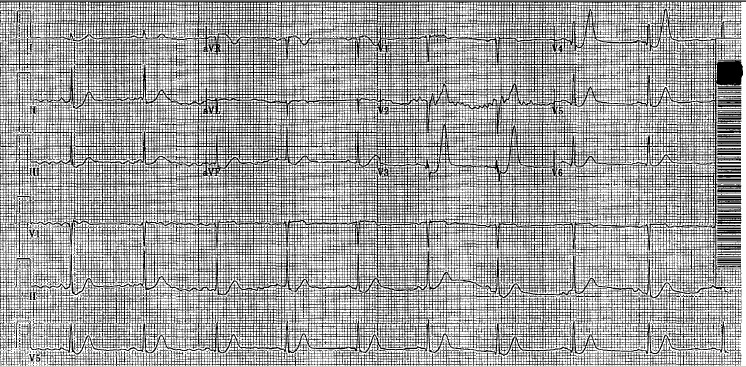
And most of the charts have a documented EKG. Ok. No problem there. Below is an example of a chart documented on a <30yoF patient with Palpitations…


Normal_ECG

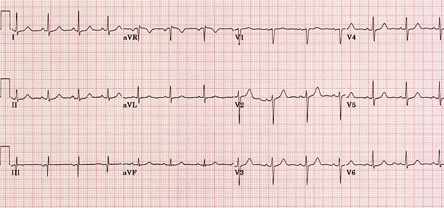
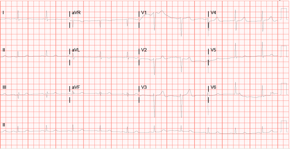
“NSR, Rate 85, Normal P waves. No S/T wave changes. Normal QTc. No ischemia. No EKG for comparison. “


The problem with this? Aside from a few parts, it is mostly irrelevant to the life or limb threatening abnormalities that we are looking for in the EKG. Sure, it will bill just fine, but I worry that there a few among us who aren’t SPECIFICALLY looking at the EKG in the palpitations patient for common causes of palpitations. I mean, really, how many of us see MIs present as palpitations?


So what should you look for in an EKG in a palpitations patient?

  1. Rate (tachy or brady or no?)
  2. Blocks
  3. HOCM
  4. Brugada
  5. AVRD
  6. Prolonged QT (cong. QT) or Short QT (why?—AVRD)
  7. WPW
  8. Ischemia


Now lets talk about the EKG abnormalities found in each:

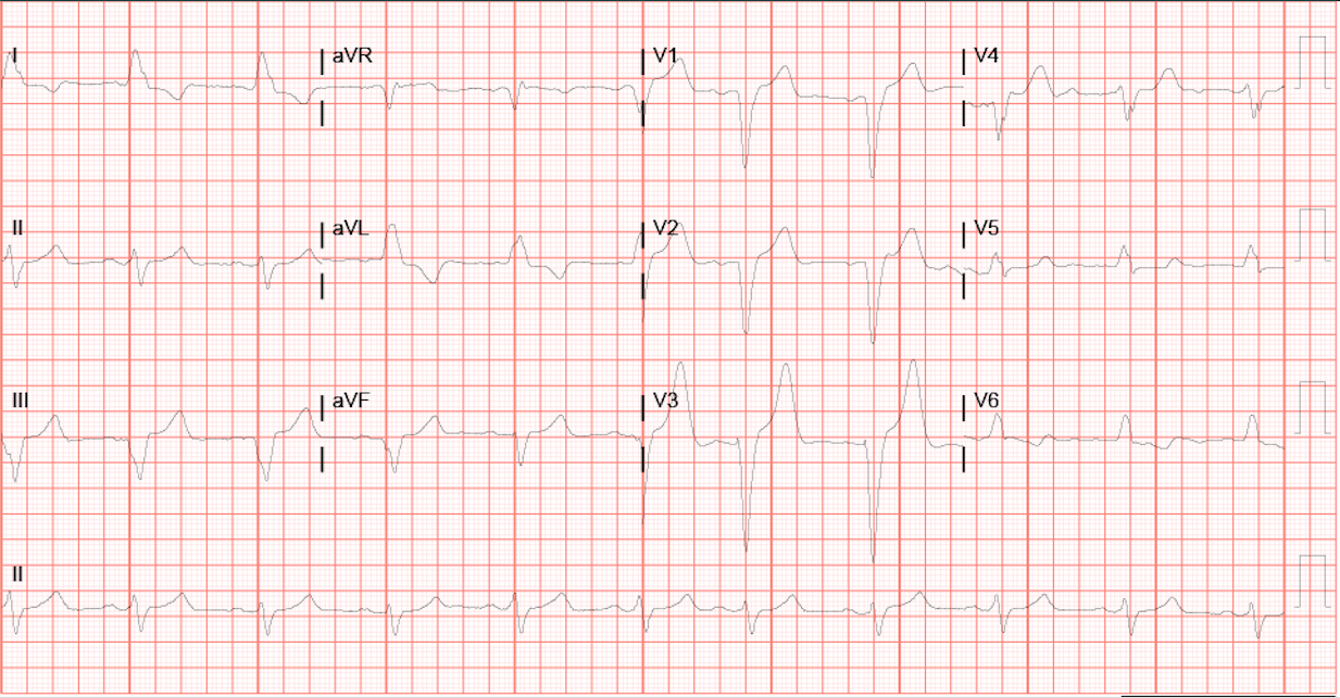
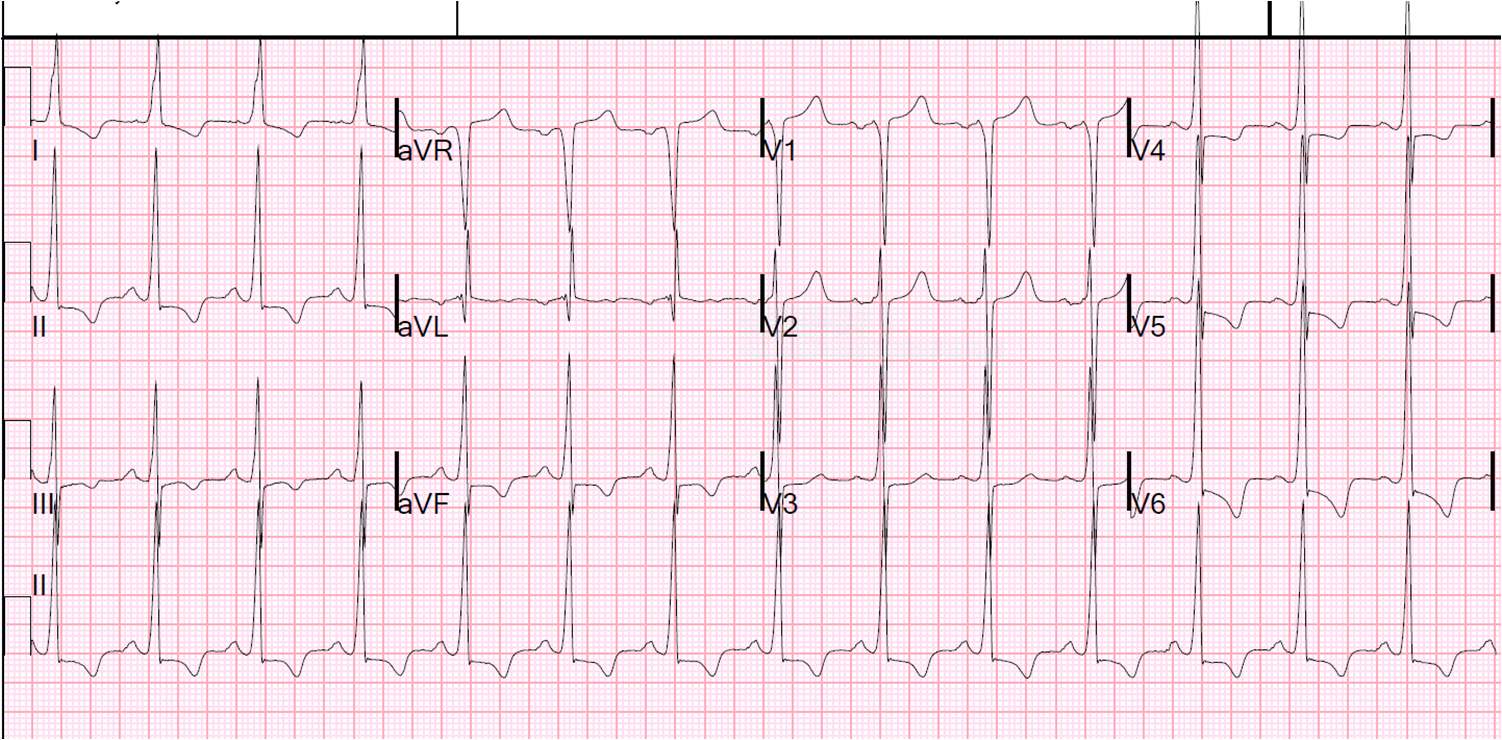
HOCM—About 90% of HOCM pts have an abnormal EKG. Remember, these are often young folks, so their ekg should be pristine. The etiology is a hypertrophic septum, so characteristic LVH is seen (V4R wave is “off the chart”), as well as deep, narrow q waves laterally. Occasionally flipped, broad T-waves are seen. There is also an apical variant that shows GIANT TWI in septal and lateral leads… you’ll know it if you see it, it looks grossly abnormal. But HOCM itself can be much more subtle. Look for Q waves and LVH then listen to their heart and refer to cardiology for an echo. Both of the below are pretty obvious, the second one is the GIANT TWI i was talking about. Not a lot of things do that.

time 4 hours HR 84

HypertrophiccardiomyopathyapicalvariantYamaguchisSyndrome

Brugada— 50% familial, most present >20yo but can be seen at birth. In brugada, you’re looking at leads V1-3, looking for a Right bundle branch block and either “coved” (st elevations with long, flat ST-segment that looks like a ski-slalom) or the “saddle type” (which is way less common and looks a lot like, well, a saddle. But remember there is STE there too which makes a side of the saddle, then the T-wave makes the other side of the saddle. This sometimes looks like a STEMI, except in a patient without chest pain and then you notice the saddle shaped concave ST segment and the CC and family history and you can smile because you didn’t activate the cath lab. Also, these patients can have EKGs that show brugada pattern SOMETIMES and not other times. Look for the ski-slope and the saddle. Hop aboard this saddle and you’re going to need a defibrillator, as anti-arrythmics don’t change mortality much….

14_minutes_QRSd_146ms_p_2_Amps_bicarb_axis_167

255v62n11-13145482fig1

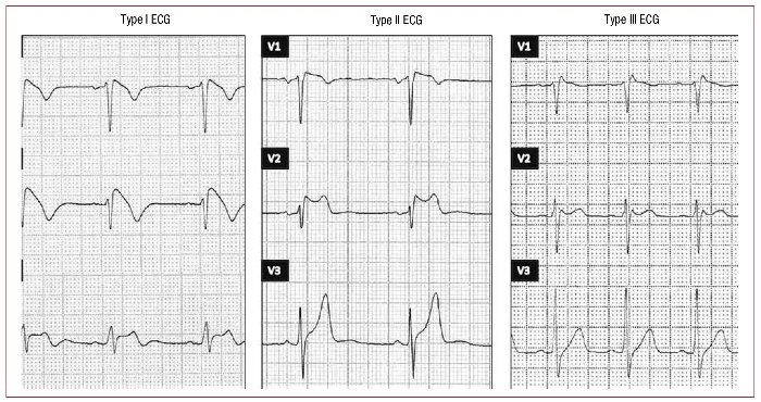
This image says there are 3 types. OK, well, that’s true and all but seriously, it’s still a saddle, so you shouldn’t be led astray.

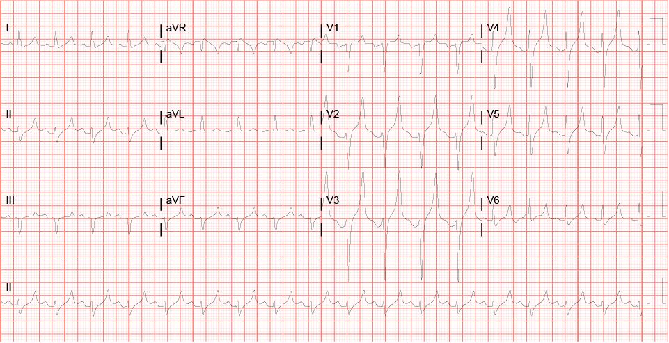
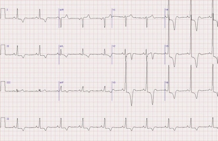
AVRD- What the what? AVRD stands for arrythmogenic right ventricular dysplasia, and basically their RV gets filled up with fat and cartilage during development. This is not so good. These folks may have exertional chest pain (it’s a cardiomyopathy). And, because it’s a structural heart disease, most of them will have EKG abnormalities (~90%). The most specific finding is an EPSILON wave, which is a little puny looking bump after the QRS that looks like a little P wave but is out of place and behind EVERY QRS. These are hard to catch, so you HAVE TO LOOK FOR IT IN PARTICULAR! Also, remember, not every Osborne wave is AVRD (is the patient hypothermic?) Occasionally this will make the QT interval short, but it isn’t reliable. Other, less specific findings are TWI in V1-3 that looks like a juvenile pattern… but remember juvenile pattern should go away when the patient becomes less juvenile. So if they have TWI in percordial leads an they are >20 and have palpitations or syncope… PANIC. No, but really, they probably have AVRD. And the syncope was probably VTACH. But they’re good now, right? Give them a defibrillator too.

avrd_figureone-2


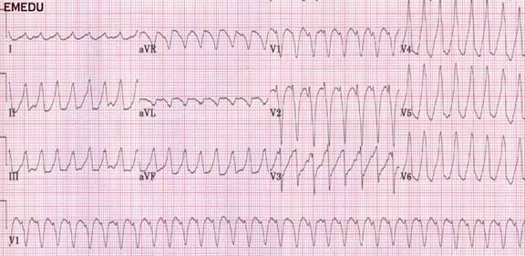
Oh, crap. That’s what happens when you send the patient home without looking for AVRD.


ARVC

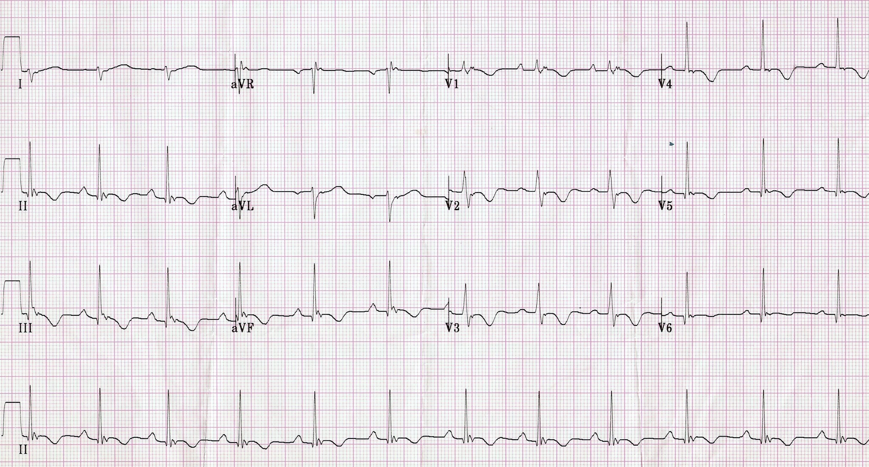
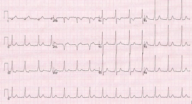
Congenital Prolonged QT-– Ok, so a bit of an update to our usual shortcut “half the R-R rule,” in Academic Emergency Medicine in October 2015, some folks published a retrospective review of some cases of EKGs with prolonged QT and non prolonged QT, and found that our quick half the RR interval thing is about 88% sensitive (r/o prolongation) but only 53% specific. Which ain’t terrible, but its also not super good (12% aren’t ruled out). Of course, what the study failed to highlight but is written and useful is that the ½ RR rule is 100% sensitive (in this study) if the HR was >60. So my take is, if they are brady, calculate it yourself, and if not, 1/2RR is good to go. Which is what I think most of us do anyway. Three cheers for studies confirming our guesses!

Screen Shot 2015-11-08 at 5.44.26 PM
WPW—Not much to say here. You know this. And you damn well should be documenting it. That PR interval is short because your patient is dying for you to find his delta wave.

Wpw

ACS– Well, this is what we do.

Blocks- P before every QRS, QRS after every P. Intervals.


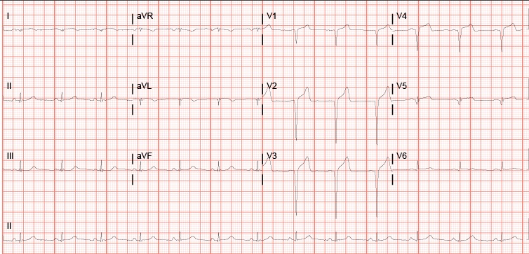
So… how should you be documenting these EKGs? With the same attention to the life and limb threatening abnormalities you are looking for on your history (early family death) and exam (murmur?). I can’t say what is right or not, but below is how I would document that EKG on our patient from earlier (NSR, rate 85, No STE/D, No TWI, no definite EKG evidence of WPW/AVRD/CongQT/blocks/HOCM).

Cheers.


Images:

https://meds.queensu.ca/central/assets/modules/ECG/Normal_ECG.bmp

http://hqmeded-ecg.blogspot.com/2014/06/history-of-hypertrophic-cardiomyopathy.html

http://www.doctorshangout.com/photo/hypertrophic-cardiomyopathy-apical-variant-yamaguchi-s-syndrome

http://hqmeded-ecg.blogspot.com/2013/05/brugada-pattern-induced-by-tricyclic.html

http://www.revespcardiol.org/imatges/255/255v62n11/grande/255v62n11-13145482fig1.jpg

http://www.geneticheartdisease.org/jpegs/avrd_figureone.jpg

http://www.heartpearls.com/wp-content/uploads/2009/07/ARVC.jpg

http://www.crkirk.com/thumbnail/arrhythmias/images/svt/ECG_WPW.htm

Another abdominal pain

I had a patient in her 30s that presented with 1 day hx of N/V and diffuse abdominal pain that was most severe in her epigastric and LUQ and radiated to her back.  She had PO intolerance since the pain started the night before. Past medical hx was significant for R nephrectomy that she states is because her “kidney wasn’t working right”. Pt says that this pain feels just like the pain she had from that kidney.

PE: VSS, afebrile. She is curled in the fetal position and yells anywhere you touch on her abdomen but states that the worst pain is when I press her epigastrum and LUQ. She has a large RUQ scar from her nephrectomy. No CVA ttp, negative murphy sign.

At this time my differential included pancreatitis vs PUD vs gastritis vs pyelonephritis
Labs come back with lipase wnl, no WBC, UA with a lot of epithelial cells and a few WBC. Acute abdominal series xray is wnl

I reassess patient after dilaudid and zofran and she states nausea has resolved but still has severe epigastric/LUQ pain. On reexamination the rest of the abdomen is nontender. The amount of pain she is experiencing in her epigastrum/LUQ concerns me and its not pancreatitis based on the lipase so I order a CT abd/pelvis and I put in the ordering comments “diffuse abdominal pain most severe in epigastric and LUQ”.

The radiologist walks over to the department to tell me that the patient has appendicitis and her appendix which is thickened and with fat stranding is in the mid right abdomen instead of RLQ hence the atypical location of her pain. My assumption is that the reason her appendix is so high is from scar tissue secondary to her transabdominal nephrectomy.

I post this to remind everyone that while the RLQ is the most specific place to have pain from appendicitis, the pain can be anywhere (previous abdominal surgery (in this case), retrocecal/pregnancy, etc).

Better to be lucky than good

I was called to a code in the ICU.  I roll into the room, 38yo IVDA hx, admitted for sepsis, septic emboli, and ARDS.   Stable vital signs 10 minutes previously, now in PEA arrest.  Already tubed and had a triple lumen in the IJ.   Not a whole lot for me to add for immediate stabilization with a secure airway and access.   Already had epi x1.  Accucheck was 149 (never, never forget this in a code).

So I immediately get to go to the second phase and start thinking reversible causes.  With that hx, all sorts of interesting diagnoses on the playing field.  The one major thing I notice was the patient was extremely cyanotic from the upper chest on up.   CBC and BMP from a few hours ago essentially unremarkable aside from a white count of 26.  Coags normal.  Actually starting to kick around the idea of empirically thrombolyzing this dude.  All of a sudden, quick run of v-fib, zap him with 200 and voila! we have a pulse.  I order a new rainbow of labs, cxr, call his pulmonologist to recite my efforts,  and strut back down stairs with a little gangster lean feeling pretty good.

I sit down, take a deep breath, and head to see the next patient.  And BOOM!  Code alarm goes off once again.  Head back into the room, next verse same as the first.  Guy suddenly went PEA arrest again, CPR was already in progress.  No labs back.  I have them pull up the CXR.

IMG_3103

Well sh*t, I swear I listened to him during the first code.  Bilateral breath sounds (course, but there).   I listen again, knowing there is a big pneumo, still can hear.  Obvious tracheal deviation on the CXR, but time to get moving.   So I ask for an angiocath and a chest tube set up.  Deer in headlights, no one moves.  Someone finally scurries off to find a pleurovac and tube.

Still waiting for an angiocath, they hand me something that looks like something I’d LP a 10 day old with, not gonna cut it.  Finally they roll in with this kit that looked like they pulled off a dusty shelf.  Has what looks like a 8 inch spinal needle with a pigail catheter already attached to it.  That’s it.  nothing else in the kit besides and adaptor to attach to the pleuravac.  Don’t have a lot of time to search for anything else so I went with it.  Was actually really slick- put the needle into the chest cavity, pulled out the inner cannula, heard the hiss and fed the pigtail.  That’s it, working chest tube in place in about the same time it would take to do a needle decompression.

IMG_3105

Immediate return of pulse and stable blood pressure.   Thank god.  That would have been a totally reversible cause of death in a young patient.  If I hadn’t ordered or remembered to check that x-ray, would have been on my shoulders.

I post this for a couple of reasons.

#1 Don’t get too cute in your codes.  Start with the basics and build from there.  I was too busy thinking about lytics etc, and had an easily reversible cause right in front of me.  Don’t forget your DOPES mnemonic for ventilated patients.

D- dislodgement, check your tubes, end tidal or even preferably direct visualization.  Especially in a patient getting CPR.

O- obstruction, always suction, mucous plugging is an easy removal.

P- Pneumo, already talked this through.

E- Equipment, I recommend always pulling your codes off the vent and bagging through the ET tube to take this one off the radar.

S- stacked breaths.  Too much PEEP or high respiratory rates without adequate expiration time can cause air trapping and decreased venous return.  Once you pull someone off the vent and bag.  Take a second and manually press the chest wall down.  Can fix the problem immediately and you look like a total ninja if it works.

Interesting case from the weekend – thoughts?

Hey guys, I was hoping to get your input on an interesting case I had at Kosair over the weekend.

16 yo F (6 ft, 150 lb…so basically an adult) with a PMH of depression, self injury, and prior suicide attempt presents after ingesting citalopram 40 mg x 90 pills (her prescription, just filled 2 days ago) and concerta 10 mg x 8-9 pills (her brother’s). Patient had been at a party the night before, admitted to EtOH.  Parents found out about the party the morning of admission and they had a big fight, took away car keys, etc. Patient decides to retaliate by swallowing pills, doesn’t tell anyone. Parents find her altered about 10:30, at Kosair at 11:40. Best guess is ingestion occurred sometime around 9-9:30am.  Had one seizure at home per family, and one en route per EMS. Generalized, tonic-clonic, brief.

Initial exam shows a drowsy but arousable patient. Answers orientation questions x3. Initial vitals show HR 147, BP 135/70, RR 25, 93% on some oxygen (can’t remember if NC or nonrebreather). Patient denies CP, palpitations, SOB, abd pain, N/V, weakness/numbness.  4mm, PERRL. MAE equally. Old self injury scars noted on wrists bilaterally. Exam otherwise unremarkable.

We start IVs, get her on a non-rebreather, get IV fluids going. Agree that charcoal seems like a bad idea with her mental status and seizures. Mom has shown up, and as we’re getting some additional history from her, respiratory is placing EKG leads. I’ve talked to poison control. Then, about 20 minutes into her stay, she seizes again. We bag her through the seizure, again generalized tonic-clonic, and just as we’re pushing 2 mg of IV Ativan she comes out of it. She appears post-ictal, but is maintaining her airway. We load her with Keppra, and as I glance at the monitor behind the attending’s head, I notice that her rhythm has changed and she looks like she’s got a wide QRS. We confirm she still has good pulses, still out of it mentally, and since she’s already connected to the EKG leads we grab one (time stamp 12:04):

EKG 1

By the time we get this printed off (!!!!) she appears to have spontaneously converted back to sinus on the monitor. But woah, holy wide QRS/long QT batman! As the attending and I are pouring over the first EKG we get another one immediately (time stamp 12:08):

EKG 2

Thankfully, the QRS appears to have normalized, but we’ve still got a loooong QT, one of the things poison control definitely told us to look out for. Having seen a few similar ingestions at University, I suggest it’s time for bicarb. The attending wants to confirm and we quickly call poison control back, they agree and suggest starting a bicarb gtt, with pH goal of 7.45-7.55.  Now we look back at the monitor and she’s throwing a ton of PVCs, captured here on EKG #3 (time stamp 12:12):

EKG 3

At this point, we opt to push an amp of bicarb while we’re waiting for pharmacy to tube up the bicarb gtt. I have to say, we see it start to work pretty darn quickly. The PVCs slow down, and her rate really starts to head back towards normal. We get an iStat (shot me down when I suggested one earlier), and a few minutes after we’ve pushed the bicarb we get an initial pH of 7.15, pCO2 of 51.5, HCO3 of 18, BE -11, and AG of 21. Electrolytes were WNL. By this time we also know her pregnancy is negative, and her serum tox is negative, no acetaminophen/salicylates on board.  At this point, we talk about intubation as the patient’s mental status is still waxing/waning and she’s breathing shallowly with brief periods of apnea, almost like an opiate overdose. Attending wants to hold off, so I go off to call the PICU resident…and end up having to hang up the conversation halfway through when he changes his mind.

So we intubate her (finally got a peds tube…in an adult), the bicarb gtt comes up from pharmacy, they’re cleaning the PICU bed her, the last EKG looks 1000x better, and all’s well that ends well (time stamp 13:08):

EKG 6

So I’m curious to see what your all’s suggestions/thoughts are on this case.  Looking back at that first EKG, how would you classify it? We’ve got a wide complex, monomorphic tachycardia that to me looks like sustained V tach (with a pulse).  The long QT doesn’t surprise me, but this rhythm does as you’d typically you’d worry about it devolving into Torsades, but that’s not what this is.

Looking back, things I would have done differently:  get a temperature sooner/order a total CK (serotonin syndrome could have been a factor and we don’t have a recorded temp until she’d almost 2 hours into her stay, no one ever ordered a CK), intubate sooner, loading her with keppra when she hit the door after 2 witnessed seizures, maybe could have prevented the 3rd?

Also, if you’re curious, I found this “Toxicology Conundrum” on LITFL that specifically discusses citalopram overdose. Has some good info, citalopram is definitely one of the more potent SSRIs, and QT prolongation is dose dependent and can be seen after ingesting >600 mg (this chick took 3.6 GRAMS). Seizures are also fairly rare, only seen in 2-3% of cases.

Intractable Pain

I’ve seen about 3 cases now that presented with intractable pain. One was leg pain, put him in Room 9 much to the dismay of nursing, dude ended up having an acute arterial clot and received an amputation. One was acute on chronic back pain (history of chronic norco use, no change in quality) that ended up with an infected ulcer on his aorta (died later). And now this case….

67 y/o M presents with hip pain. Reports he was riding on a mower, hit a bump, had mild pain afterwards in his hip, but overall did well. He ambulated inside, took a shower, and after he sat down to do his business on the toilet he started to worsen. Especially when he stood up. No numbness/tingling/weakness/nausea/vomiting/fevers/chills/bowel or bladder incontinence. VSS. He has pin point tenderness behind his R hip. Worse with palpation. NO pain with axial load, internal/external rotation of leg. Pain with movement of torso. Pain isn’t nearly as bad when he’s just laying there. Appears in pain though at all times. Like TOO much pain.

Skip the XR knowing it won’t satisfy me, go straight to CT pelvis w/o contrast. It is normal. Decide to check labs at this point because I forsee an admission. He has a WBC count of 12. Cr of 1.8 (which is old) I admit him to the hospital for pain control as the idea of ambulation wasn’t happening, let alone just sitting up in the bed.

He is discharged the next day after MRIs of the L spine and hip are showing degenerative changes and lumbar stenosis and he is walking now (with the assistance of PT)

He presents again today and sees one of my partners. Again appears in pain, uncomfortable. Now has abdominal pain in the LLQ as well CT shows DIFFUSE pneumoperitoneum. He’s in the operating room at this moment…..

Sorting out theatrics/drama from reality can be a fine line we walk. Opiate abuse complicates things, and we often meet plenty of actors. But keep in mind pain out or proportion is almost always bad and delineating that can be difficult!

Always have your radar up, don’t be afraid to work up patients (regardless of what your ancillary staff and colleagues say)….. and don’t be afraid to be patient advocates and put them in the hospital to allow things to develop…..Remember One EKG, one CT, one lab draw, they are all stand still pictures of one moment in time and don’t always tell the whole truth. Certainly didn’t here in this case…..

EKG Changes in Hyperkalemia

I have had a couple of good EKG’s in the setting of hyperkalemia that I thought I would share.

The first case is a 68 y/o female with ESRD on dialysis presenting with “back pain”.  Turns out that her back pain was actually chronic and at baseline, but she had missed her last two dialysis appointments.  She denied any chest pain or SOA.  I ended up getting an EKG while waiting on labs.

Initial EKG:

Screen Shot 2015-08-04 at 2.46.53 PM

Previous EKG (~6 months ago)

Feb


What says you?  New onset LBBB?  After seeing this we gave Calcium Gluconate and asked one of our wonderful techs to grab a quick iStat as labs were taking forever to result.  Potassium was 6.8.  Gave insulin + D50 and bicarb.  EKG was repeated after Hyperkalemia treatment:

post

Renal consulted for emergent dialysis and medicine admitted.


Here’s another EKG that I had recently from a DKA patient who had an initial potassium of 7.8:

Hyper-K


Here’s a couple of good hyperkalemia resources:

Hyperkalemia EKG Basics

Treatment of Hyperkalemia

Snakebite

Recently saw at case at ULH (Not mine but posted with permission from resident involved) of a young female (18-20yo) flown from OSH for copperhead bite to L foot. Pt had been hiking in the woods with her boyfriend when she felt a stabbing sensation on her foot, looked down and there was a snake. Pt took a picture of it – sure enough it was a copperhead. Labs at OSH wnl, sent to ULH for concern for possible need for antivenin. I realized – being an urban trauma center – we don’t see a lot of snake bites. If you’re from the area, you may not be familiar with our venomous critters.
There are four species of venomous snakes in KY, all pit vipers (triangular heads, heavy bodies, cat-like pupils) – fw.ky.gov
1. Copperhead – extremely common throughout the state, most common envenomation, mildest venom of the four. (As an acquaintance of mine, Jim Harrison of East KY Venom Extraction lab used to say – no recorded history of death from copperhead envenomation in KY history
2. Timber Rattlesnake – found throughout state, potent venom, relatively docile. Dark coloration, have a rattle (obviously)
3. Pygmy Rattlesnake – found in extreme Western KY. Also have a rattle, potent venom
4. Cottonmouth – found in Western KY, potent venom, can be aggressive. Can be mistaken for Eastern Water Snake (common, non-venomous, aggressive water-dwelling snake found throughout KY)
Best identification – hopefully someone took a picture – otherwise, one to two small puncture wounds with increasing swelling and pain are good signs of envenomation. However, approximately 25% of viper envenomations can be dry bites – where snake gives a warning bite, injects no venom.

Signs and Symptoms of snake bite:
Venomous snakes in KY typically have venoms containing cytotoxins and hemotoxins – they break down tissue and can act on coagulations factors
-puncture wounds (usually paired, can be one)
-erythema and swelling
-intense pain at site and increasing proximally
-systemic symptoms can include nausea, vomiting, abdominal pain, vertigo. Altered mental status and hypotension can present as well

First Aid if bitten:
-Walk slowly – increased activity increases circulation of venom
-Elevate affected part: most bites are on distal extremities: feet, hands. More serious complications are associated with bites involving trunk, neck, face
-Wash with soap and water
DO NOT:
-Apply a tourniquet
-Attempt to suck venom from wound
-Apply ice to wound

Medical Work-up for known or suspected envenomation
-ABCs (always the first step)
-Good H&P – observe amount of swelling, movement of affected area. Mark swelling and erythema with marker to observe for progression
-CBC
-Comp
-Coags
-Total CK
-Fibrinogen
-U/A – to look for myoglobinuria
-Consider ABG and lactic acid if signs of systemic toxicity
-Xray to r/o retained teeth

Severe complications
-Rhabdomyolysis – from muscle breakdown from venom
-Compartment syndrome – cannot be diagnosed on clinical exam alone. Venom can cause tingling and paresthesias. If concerned, consult your surgeon
-Thrombocytopenia and coagulopathy – caused by thrombin-like molecules in venom. Treated with antivenom
-Systemic toxicity – can be life-threatening

Treatment:
-Update Tdap if indicated
-Pain control, elevation
-Antivenom if indicated

Antivenin: Always contact your toxicologist prior to giving
-Recommended for moderate to severe envenomations
(http://www.uptodate.com.echo.louisville.edu/contents/image?imageKey=EM%2F53948&topicKey=EM%2F6595&rank=2%7E39&source=see_link&search=snake+bite&utdPopup=true)
-Dosing: Moderate envenomation – 4 to 6 vials (1g each) over 60 min with repeat dose as needed
Severe envenomation – 6 to 9 vials over 60min with repeat dose prn
-Contraindications: papaya allergy (contains enzyme papain)
-Stop if signs of anaphylaxis

Disposition
-Requires antivenom – admission. Complications can include compartment syndrome, delayed coagulopathy (up to 48hrs). May need redosing of antivenom
-Mild envenomation or no symptoms – can be discharged home if pain controlled and no signs of toxicity at 8-12hrs s/p bite. If bitten by rattlesnake or cottonmouth, recommend CBC, PT, fibrinogen at 2-3d and 5-6d after initial bite

A large red herring

The patient is a 20s y/o AAF, multip, last normal period 3 weeks ago (definite date), who presented to another facility with spotting and occasional moderate bleeding for about a week with 4 days of crampy pelvic pain, worse on the left than the right.  There, she was diagnosed with trichomonas and treated empirically for gonorrhea and chlamydia.  She had a positive pregnancy test and reportedly had transabdominal and transvaginal ultrasounds.  She stated they told her she definitely had a pregnancy in her left tube.  She was given instructions to see her regular doctor in 48 hours for a repeat evaluation and discharged.

It is at this point that the big red flashing lights in my head go off and I think to myself “Wait, WHAT?  Someone DISCHARGED a confirmed ectopic?  No way.  Maybe they said it was a possibility and she misinterpreted.”  Whatever the sequence of events, all that mattered was that I get the records and do an ultrasound myself.

Her records came in from the other ED after a couple of attempts (as per usual).  A few things that I learned from her records:
1. she was indeed told that she had an ectopic and that she should see her regular doctor or OB in 48 hours.
2. she did get treated for STDs.
3. sure, she had an ultrasound, but there wasn’t any interpretation available unless I called and had them hold the phone up to the speaker (didn’t have time to do this).
4. my dislike for paper charts is warranted when I can’t read what someone says about my patient.

Initial vitals normal with a HR 100, BP 118/78
She was tender in the LLQ and suprapubic areas without peritoneal signs.  Cervix was closed, she had a small to moderate amount of dark blood in the vaginal vault, and had uterine and left adnexal tenderness on bimanual exam.

I started fluids, gave her some meds for her nausea, and put in all the usual orders.

Labs:
-WBC 13.8 (88% Neut)
-Hb 9.6
-CMP unremarkable
-U/A positive for blood/protein/ketones but micro negative
-Quant 12000+
-Swabs + for trich

And then came the ultrasound:

transverse view, abdominal probe

transverse view, abdominal probe

longitudinal view, abdominal probe

longitudinal view, abdominal probe

A few things about this caused even more red light flashing: first, how could a gestational sac be THAT SIZE in someone whose last period was 3 weeks ago?  Second, what was that OTHER thing behind the uterus on the left?  Third, if the sac was really that big, why was there no pole?

The transvaginal exam was more or less to confirm my suspicions that this was not normal.  I knew that my plan was now to call the specialists with the fancy machine and adnexal expertise.  I couldn’t actually get a GOOD view in two planes (sorry, Dr. O’Brien!) due to patient discomfort, but this one was good enough.

longitudinal view, transvaginal probe

longitudinal view, transvaginal probe

I hadn’t seen any free fluid in her pelvis on either exam.  However, when OB came down, they found some.  They confirmed my suspicions and admitted the patient to go to the OR.

In my brief lit search just prior to posting, I found that this pseudosac finding is not extremely common (the average reported frequency is about 10% of cases).  I feel like this patient’s story would have raised enough red flags to make me uncomfortable sending her home without OB involvement even without the ultrasound, but the date/quant discrepancy coupled with a sac that was definitely not consistent (even though it WAS in the uterus) clinched the diagnosis for me.

RLQ pain and N/V

15 yr male with hx of hemophilia presenting with 1 day hx of progressively worsening RLQ pain, decreased PO, nausea, and vomiting. Described RLQ as a “small swelling’ that continued to span across the R abdomen as the day progressed. Denies dysuria, hematuria, hematemesis, hematochezia, constipation, diarrhea, abd trauma, or testicular pain. No previous abdominal surgeries. Physical exam is significant for RUQ and RLQ tenderness, no obvious swelling, no ecchymosis seen. He definitely appeared ill and uncomfortable. A&Ox4.

So already…what are we considering?  Appendicitis …. Peritoneal bleed … bowel obstruction …maybe a few others (UTI, Kidney Stones, STI).

While waiting on CT Abd/Pelvis imaging to be completed, patient is found to be anemic with a Hgb of 8. Normal WBCs. Platelets: 300. Elevated PTT: 83. Normal PT/INR. Urinalysis…. negative. IV Fluids have already been started. Zofran for his continued nausea.

Here’s a significant snapshot of the CT

Abdomen

———————

It spanned from the R kidney down to the bladder. Actively extravasating. Hydronephrosis due to the hematoma compressing the R ureter. It compressed the R renal vasculature as well, and anteriorly displaced the R kidney.

Contacted Hematology, where we decided to administer FEIBA. (He usually takes Alphanate MWF, but had not taken any medicine on day of presentation. Plus, the hospital did not have his particular medication, so we needed to find an alternative.) He was admitted to the Hematology service. They have plans of contacting Surgery for any possible interventions once his Hgb stabilized.

Repeat CBC (after patient had been admitted) showed that the Hgb had fallen to 6.0.

Diagnosis: 15 year old male with non-traumatic R retro peritoneal hematoma. Source currently unknown.

A-fib with RVR + sepsis + hypotension = conundrum

How do we slow the rate without lowering the BP? Will slowing the rate even help the BP? How do we raise the BP without speeding up the rate?

The patient who inspired this post came in for a bowel obstruction. Cards was initially consulted for possible new a-fib, but there really wasn’t much to do from a cardiac standpoint. THEN he perforated his bowel and went for emergency surgery, where he required pressors and went into a-fib with RVR. He was packed open and taken to the SICU, where he was hypotensive to SBP 70s, tachycardic to 160s, and intermittently hypoxic.

If you want to skip my thoughts on the case and head straight for the facts, here is an interesting article about a-fib in critically ill patients. It talks about the various options for management and the pros/cons of each.

If expert opinion is more your style, try this.

Of course, there is also controversy around slowing down a-fib when it is caused by sickness. Should we let the body do its thing to try and compensate? Here’s one article that suggests maybe we shouldn’t get so hung up on rate control in sick people.

Now back to the case. The surgery resident and I were of the mind that slowing the rate and organizing the rhythm should help with cardiac output. There are several reasons why this logic still seems to be in the majority.

– With a-fib you lose atrial kick. That little extra oomph from the atrium may not seem like a big deal, but it can have a significant impact on cardiac output. Here’s a fun article from 1965 that shows a 53% increase in cardiac output from converting a-fib to sinus (using quinidine because 1965). The results have obviously been redemonstrated in more recent studies, but how often do you get to reference quinidine? Not that often.

– Diastolic filling is important for stroke volume. With any tachyarrhythmia, less diastolic filling time means lower stroke volume. However people who do “math” would argue that increasing the rate would likely keep cardiac output about the same. This logic holds up with regular rhythms, but studies show that irregular rhythm decreases cardiac output compared to regular rhythm. Here’s one such study.

– In real life, we went for amiodarone and electrical cardioversion. From the a-fib in critical care article above, it seems like that’s still the best option.
Here’s a good article I wish I had read before embarking on a cluster of a cardioversion. P.S. Put the pads on correctly. Anterior-posterior pads definitely worked better for this 350 pound patient. Anterior pads = fail x3. AP pads = success!

– It’s worth noting that this is not one of the scenarios when you’re worrying about giving someone a stroke with cardioversion. This guy’s risk of death was much greater than his risk of stroke.

Another consideration for this situation was the choice of pressors. Eventually the patient ended up maxed on pretty much all pressors, but that may not always be the case.

Surviving Sepsis guidelines are all about Levophed as a first line pressor, which is usually a great option. But guidelines are just guidelines. How many of our patients are otherwise 100% healthy and just have a little sepsis? Not that many.

– In this case, I think phenylephrine may have been a better first option. Pure alpha agonist activity vasoconstricts without Levophed’s cardiac effects, which probably didn’t do us any favors with the RVR.

– Based on the EMCrit blog above it also seems like phenylephrine might have allowed us to use a beta blocker without worrying about blocking the effects of the pressor or a calcium channel blocker without exacerbating hypotension. Thoughts?

– Anyone have other ideas about pressors in this scenario?

There was a lot more to the case after that, but this post has already ended up way longer than I intended. In the end, it was a 350 pound unhealthy guy with a less than ideal heart, so unfortunately, his family ended up withdrawing care. I doubt anything we could have done would have changed his outcome, but maybe there’s something to learn from it that will help someone else someday. I’ve talked to several people about this case and gotten different opinions from each one, so I figure why not open it up for a few more opinions to really confuse clear things up.

Everything including the lipid sink

80+ yo gentleman with PMH of COPD, CHF, CAD, CKD, initially hospitalized for urosepsis and NSTEMI with a worsening AKI, and baseline CKD. Tuned him up and sent him to the floor where he accidentally received an overdose of his carvedilol. Got the call that the patient was hypotensive to 70s systolic, bradycardic to 30s.

Immediately pacer pads were placed to ward off evil spirits
-Started with 1L crystalloid bolus –> no response and with risk factors and pmhx, didn’t want to fluid overload
-Tried atropine (3-4 doses) –> no response
-Tried glucagon 50mcg/kg loading dose followed by 3mg/hr infusion –> still no luck and exhausted all the glucagon in the pharmacy
Running out of options here. I remember reading an article in Annals about using lipid infusions in beta blocker overdoses and figured it was worth a shot.

Started 20% Intralipid bolus through central line at 1.5ml/kg followed by 0.25ml/kg/min for 30min. Maybe it was the lipid infusion or just the combined effects of everything but by 6am, pt’s BP and HR had improved to normal limits and he began talking again.
It was a nice result but one I couldn’t celebrate that much – within a week, patient decided to go to palliative care and that was the last I heard of him.

PEM Chronicles: Rasburicase

     A case I saw  last month led me to utilizing a drug I’d never heard of before in the ED: Rasburicase.

     The drug: a recombinant urate oxidase enzyme, which converts existing uric acid to allantoin. This is key due to the higher solubility of allantoin in urine. Patients with Tumor Lysis Syndrome are at risk of acute renal failure due to precipitation of uric acid crystals in renal tubules and collecting ducts.

The case: a 12 yo F with no PMHx who was transferred from an urgent care center for multiple tender, enlarged lymph nodes and a WBC count of 98.

As we worked her up for a new presentation of a hematologic malignancy, it became evident she fit into the parameters for TLS. Initial uric acid level – 14.5 along with hyperkalemia, elevated serum LDH, and hyperphosphatemia. When Hem/Onc came on board, once labs were back, the first recommendations were hydration and Rasburicase.

Provided below is the article I found that informed me on the treatment and a few key points picked up when using it.

For kids only – The drug hasn’t been approved in the US for adults.

G6PD deficiency – Yes, from the depths of med school knowledge, this condition is a contraindication to using Rasburicase. The hydrogen peroxide it produces as a byproduct can lead to hemolysis. Ask if there is a family history.

Coordinating with pharmacy – Due to the preparation of rasburicase, it should be administered immediately after it has been prepared. IV access should be obtained well in advance.

Type of IV access – In our case, hem/onc was considering emergent dialysis. It may be prudent to discuss this with your consultants to get a head-start on what they’ll need once going upstairs.

Not at your medical facility, a specialized pediatric hospital, or somewhere with this in stock??? As you transfer, consider:

1.) Aggressive IV Hydration. Easy to start getting this started, especially if transporting to the next hospital will require hours instead of blocks.

2.) Allopurinol. Though it isn’t required when Rasburicase is given, allopurinol can’t decrease what is already present, but it can help prevent the formation of more uric acid.

http://www.ncbi.nlm.nih.gov/pmc/articles/PMC1200736/

The elusive S1Q3T3

So this is a case I thought was interesting that I had in the department back in May. We all know that the most common EKG finding in the setting of PE is sinus tach, however the pimp question that is also asked is the finding of S1Q3T3. While I can easily recite the alphabetic-numeric code S1Q3T3 by heart at the drop of a hat, I had never seen one and honestly thought I never would. I thought finding S1Q3T3 was likely as rare as surviving a ED thoracotomy (OK maybe not that rare).  So on to the case.

39 year old female presents to the ED complaining of SOA and cough for the past two weeks. Cough was productive of green sputum, no fevers, and she does complain of some chest pain which sounds pleuritic in nature. Initial vitals HR 118 BP 111/73 RR 16 O2 97% room air. As I get into her PMH she says she has a history of multiple PEs with an IVC filter placed 1 yr ago because apparently she wasn’t very good at remembering to take her coumadin. She has had a hypercoagulability workup which was negative, no recent travel, no estrogen use.

So at this point with a history of multiple PEs, tachycardia, SOA, and pleuritic chest pain I am thinking I am going to scan this lady. Even though she had a IVC placed a year ago, she is still saying all the right things for PE. So while the CTPE protocol was cooking I got an EKG and there it was, S1Q3T3!

S1Q3T3

Needless to say I was pretty excited and immediately showed the rotating intern next to me who clearly didn’t share my enthusiasm. When I compared this new EKG to a past EKG a month ago she did not have the S1Q3T3.During her admission a month ago, when she had a normal EKG, she had a CTPE showing a chronic PE. This time when her CTPE came back the read was Acute on Chronic Pulmonary Embolism. So a month ago she had a chronic PE with a normal EKG and at this visit she had an EKG with S1Q3T3 and a acute on chronic PE. Out of curiosity I dug through her medical history a little bit more and found that this patient had multiple prior admissions for PE with multiple CTPE protocols and EKGs. What I found was that whenever this patient had a CT read of Chronic PE she had a normal sinus rhythm EKG. However, whenever she had a read of Acute on Chronic PE (which was 4 times!) she had a EKG showing S1Q3T3, dating all the way back to 2012. Yea, apparently this lady has been hanging out with a chronic PE in her distal right main pulmonary artery since 2012 and every once in a while she will throw a new small PE, even with an IVC filter.

So after doing a little bit of thinking and a little bit of reading it made sense. The EKG finding of S1Q3T3 is indicative of right heart strain, in this case resulting from an acute PE. So this patient’s heart has adapted to her chronic PE, however every time she throws a new PE she has an element of right heart strain which can be seen on her EKG as S1Q3T3.

I just thought this was pretty interesting to actually see the physiologic and mechanical adaption and strain this patient’s heart was undergoing being clearly demonstrated on her EKG. Also I learned that S1Q3T3 is not like a q wave after an MI in that it stays on the patient’s EKG, it is a finding that comes and goes depending on the patient’s presentation.

Anyways I thought this was a pretty cool little case and figured I would share. Hope you all enjoyed.

The Kitchen Sink

Fairly early into an overnight shift the radio goes off.  EMS is approximately 6min out with mid 30s male undergoing CPR. Per bystanders, the patient was going running from door to door, banging on doors and yelling for help. Shortly after being taken into police custody he was found to be pulseless. Had been undergoing CPR approx 10 min. King airway in place. No improvement with dextrose and naloxone.

The intern on and myself go to Room 9 to get set up. When EMS arrives about 10 minutes later, CPR is still ongoing and rhythm has been asytole/PEA throughout. The story remains consistent with him going from door to door yelling for help but now there is some variation as to whether he was cuffed and then lost his pulse, was found without a pulse, or if he got into a physical altercation with the neighbor and then lost pulse shortly after arrest. There are no signs of trauma and we elected to not invite additional chaos by calling a Level 1.

CPR is continued. King airway switched out for ETT. Accucheck 250’s. Central venous access and bilateral chest tubes placed with no rush of air or blood. Sats persist at 85% on vent, etco2 is in 60’s. He gets epi q3m. A dose of vasopressin and steroids. Istat has K 5.5. Lactate >20. Received 2-3 amps of bicarb. Received calcium. Upon rhythm checks he was primarily asystole except for 2-3 checks with a narrow pea that would not persist. Each time the ultrasound was placed on his heart there was no cardiac activity. My thought process at this time was that he was either excited delirium that I’m not going to be able to do much about or some kind of ingestion/overdose. We called pharmacy and asked for lipid emulsion. His ETCO2 had remained between 40-60 and we were about 40 minutes out from reported time of arrest. I decided if the lipids were unsuccessful that would be when we would call it.

They arrived about 15 minutes later and were infused. By now his etco2 had drifted down to 10 on the most recent pulse check. No changes occurred and TOD was called.

Reasons not to get into prison fights…

Middle aged male transferred from an OSH, accepted by ENT for a mandible fracture.

The patient is incarcerated, and was involved in an “altercation” with other inmates. The incident occurred around 2PM; but he didn’t report any of his pain to the guards until 10PM.  On arrival at the OSH he had multiple contusions to his face/head, lacerations over his hands, and obvious dental trauma.  The patient was also complaining of chest pain – he stated that another inmate had slammed him in the chest with his knee. Despite his age, the patient has a history of previous MI in 2011, cathed at U of L with no stents placed. Takes a baby aspirin, no other meds and no other PMH.

At this point, the patient is about 10 hours out from the incident. Work-up at the OSH with the following: neg CT head and CXR. CT face with a mandible fracture. Labs notable for WBC 17.8, Hgb 14.3, platelet 373, normal coags, normal electrolytes, BUN/Cr 14.0/1.1. Total CK 213 (55-170 normal), troponin <0.012, CKMB  1.66 (0 – 3.38 normal), myoglobin 271.8 (0-121 normal).  Tox screen negative. EKG is as follows:

OSH EKG

His hand lacerations were repaired and he was started on Augmentin for a human bite. ENT accepted, and the patient was transferred to U of L, arriving about 6 AM. Dental was consulted on arrival and splinted his teeth. By 9 AM ENT had evaluated the patient and admitted him to the floor, planning for surgical intervention.

The patient was an ED floor hold, and around 2PM began complaining of worsening chest pain. ENT was paged and ordered an EKG and a set of cardiac enzymes, coming down to re-eval the patient. His EKG now looked like this:

1410 EKG

Enzymes came back with CK total 5024, CKMB 303, and troponin 44.1. Cardiology was consulted and ordered a stat echo and started the patient on ACS protocol. The echo showed an EF of 30%, an akinetic mid/distal anferoseptum and an akinetic apex. Cards initially thought that this was consistent with stress cardiomyopathy in the setting of trauma, but couldn’t rule out cardiac ischemia due to direct cardiac trauma. They planned to treat medically and cath in the morning.

Throughout the evening, he developed worsening ST elevation in his lateral leads and his troponin continued to rise, up to 67.0 by midnight.

0308 EKG

The on call cath attending at Jewish was consulted and by about 3AM the decision was made to transfer the patient to Jewish for a cath first thing in the morning.

Final result: 100% LAD occlusion, secondary to direct cardiac trauma.

Definitely rare injury, but one to keep in the back of your mind, especially as it can occur in previously healthy, relatively young patients. Of note, these can have delayed presentations, up to several days. Typically occur after MVA, but there are several cases reports occurring after crush injuries, being hit in the chest by a soccer/rugby ball, and my personal favorite, one listed as “struck in the chest by an umbrella tip.”