PPE Videos

In case any of you need a refresher on donning/doffing PPE, see videos below. The first is on normal PPE. Would recommend skipping to the 5 minute mark or so on the first one.

https://nam03.safelinks.protection.outlook.com/?url=https%3A%2F%2Fwww.youtube.com%2Fwatch%3Fv%3DbG6zISnenPg%26feature%3Dshare_email&data=02%7C01%7Cadam.ross%40louisville.edu%7Cb26b5cd106bc410bc36608d7ce7a6b82%7Cdd246e4a54344e158ae391ad9797b209%7C0%7C0%7C637204897498164783&sdata=src2WeSc06Os21NKeWiSnUC2U5u60ZSoJ37dIzHYNvo%3D&reserved=0

This video below is on the donning/doffing of PAPRs/hoods. We haven’t moved towards using these just yet, and I’ll provide some additional review/education/training in the near future, especially if we begin to push towards using them. Can skip to about the 4 minute mark on this one if you like.

CT Scans-Covid

First off, it’s not currently recommended to perform CT Chest on all suspected Covid patients.

If you’re doing a CT Chest for rule-out Covid-19 you must put this in the order comments/indication. If you’re scanning a patient who is under isolation precautions due to possible Covid, and order a CT PE Protocol, put Possible Covid, Rule-Out PE. The reason behind this is they’ll take the patient to the basement so that they don’t have to shut down the ED scanner. Thanks.

Journal Club January 2020

We had our January journal club at Vines, discussing 3 articles on different topics.

1- The first was an editorial on legal cases related to use of medical stents. The authors highlight the inter-rater difference in assessment of coronary blockages between even advanced interventional cardiologists. This has legal implications as cardiologists are prosecuted for unnecessary procedures. This article is important to EM no as much for the medical content (we don’t place stents), but for the precedent of the legal system prosecuting physicians for doing what they were trained to do.

2- The second article in Annals EM looked at 24,459 ED patients with chest pain who were deemed to require outpatient stress testing. The conclusion: “Less than one third of patients completed outpatient stress testing within the guideline-recommended 3 days after initial evaluation. More important, the low adverse event rates suggest that selective outpatient stress testing is safe. In this cohort of patients selected for outpatient cardiac stress testing in a well-integrated health system, there does not appear to be any associated benefit of stress testing within 3 days, nor within 30 days, compared with those who never received testing at all. The lack of benefit of obtaining timely testing, in combination with low rates of objective adverse events, may warrant reassessment of the current guidelines.” Notice that this does not change guidelines, but ads to the conversation on proper use of resources and how aggressively we should work up chest pain in low risk patients.

3- The final article, the 2018 flu guidelines, sound similar to prior years, with focus of testing and treatment on patients at risk for complications. They do throw in the recommendation to test “if the results might influence antiviral treatment decisions or reduce use of unnecessary antibiotics, further diagnostic testing, and time in the emergency department, or if the results might influence antiviral treatment or chemoprophylaxis decisions for high-risk household contacts.” Physicians should start treatment right away for

  • Persons of any age who are hospitalized with influenza, regardless of illness duration prior to hospitalization (A-II).
  • Outpatients of any age with severe or progressive illness, regardless of illness duration (A-III).
  • Outpatients who are at high risk of complications from influenza, including those with chronic medical conditions and immunocompromised patients (A-II).
  • Children younger than 2 years and adults ≥65 years (A-III).
  • Pregnant women and those within 2 weeks postpartum (A-III).

And we can consider treatment for

  • Outpatients with illness onset ≤2 days before presentation (C-I).
  • Symptomatic outpatients who are household contacts of persons who are at high risk of developing complications from influenza, particularly those who are severely immunocompromised (C-III).
  • Symptomatic healthcare providers who care for patients who are at high risk of developing complications from influenza, particularly those who are severely immunocompromised (C-III).

Hope everyone enjoyed Journal Club, looking forward to the February articles

Congestive heart failure exacerbation management

Most of the third years, and probably the second years at this point, know pretty well how to manage CHF exacerbations. However, there are differences in managing the normotensive vs. hypertensive vs. hypotensive exacerbation, and I’ll try to provide some tricks in managing your run-of-the-mill CHF exacerbation as well (credits to Amal Mattu and Scott Weingart).


First, try to determine the cause of the exacerbation. Determining the cause of the exacerbation, however, can be difficult – most patients aren’t honest about their salt/food/drug intake or medication compliance, or do not understand their disease process. The most common cause of an exacerbation is dietary or medication non-compliance. However, in all cases, ischemia needs to be considered. More rare cases can be valvular dysfunction such as rupture (auscultation or BSUS), myocarditis (check a troponin if it fits the clinical scenario), arrhythmias (check HR and an EKG), hypertensive crisis (often secondary to medication and dietary non-compliance, or drugs), or high-output failure (such as anemia, sepsis, AV-fistula in a dialysis patient, thyrotoxicosis). (1)


Next comes diagnostics. There are debates on whether troponins and BNPs need to be ordered on every CHF exacerbation. For example, many of these patients will have chronically elevated troponins and BNPs, and you’ll be stuck trending them. A BNP is of limited value in the ER except in maybe determining is the patient’s dyspnea secondary to a COPD exacerbation or CHF exacerbation, or if the BNP is normal then it should make you consider alternative diagnoses. Amal Mattu suggests that if you’re going to admit the patient, get everything (CBC, CMP, BNP, trop, EKG, CXR) because the inpatient teams like to trend trops/BNPs (even if the evidence doesn’t really back up trending BNPs). If it’s simply diet or medication non-compliance, you might only need to check electrolytes, because diuretics can cause electrolyte disturbances. ABGs aren’t typically helpful unless the patient is hypoxemic. Overall, not much other testing needs to be done for your non-critically ill patients. (1)


As for management, this becomes tricky because it depends on many things. What is the patient’s blood pressure? What is the cause of their exacerbation? Are they volume-overloaded, euvolemic, or dry? Are they septic? In general you have three goals: decrease the preload, decrease the afterload, and (in some cases) increase LV function. PLEASE watch this Amal Mattu lecture and you will master CHF exacerbations (2). For management, let’s start with the classic hypertensive patient.


Hypertensive patient: The patient’s heart is straining against a high amount of SVR and can’t perfuse their kidneys. Fluid backs up into the lungs. Don’t immediately jump to your loop diuretics (it’s debatable, and should only be done if hyper or maybe euvolemic). We fix this problem by fixing two problem: decreasing the SVR in order to perfuse the kidneys, and decreasing preload to “turn off the faucet” that’s overflowing a bathtub (lungs). Lasix won’t work if you aren’t perfusing the kidneys. One of the best and quickest agents we have is nitroglycerin. You have three options: 0.4mg sublingual tab, nitro paste, and a nitro bolus and drip. Scott Weingart (3) recommends the nitro drip, starting with a bolus of 400 mcg (that’s one sublingual nitro tab) over 2 minutes, and then drop to 100 mcg/min and titrate up until SVR is decreased. Your nurses will often question if you want to start the rate that high, and the answer is yes, but you MUST monitor your patient closely because high doses of nitro can tank your patient’s BP if you aren’t careful. Also, nitro also decreases preload, which can prevent fluid from backing up into the heart and therefore the lungs. (Nitro often gives patients a headache – give ‘em some Tylenol.) This can fix patients in minutes, because you’re redistributing fluid out of the lungs and into the vasculature or the rest of the body! BiPAP will save these patients by pushing fluid out of their lungs and can prevent intubation. The other thing to consider is IV ACE Inhibitors. Enalapril (enalaprilat) is the only IV form we have, and it has to come from pharmacy, but ACE Inhibitors are very good at afterload reduction and will help move the fluid from the lungs into the rest of the body. If nitro isn’t working, consider adding on enalapril.


Normotensive patients: just because their BP isn’t high doesn’t mean you can’t use nitro. However, be judicious, because you don’t want to cause hypotension, but remember that even normotensive patients can tolerate a sublingual nitro without issue. Lasix if clinically volume overloaded. BiPAP as needed for respiratory difficulties. Otherwise, nothing too tricky here.


Hypotensive patients: these are actually your cardiogenic shock patients. They are sick as snot. They could also be septic (! Hi Dr. Shoff), or could be having an MI. Your management of these patients is very difficult and much different than your typical CHF exacerbation management. First, determine if the patient is “warm” or “cold” (feel their extremities), and then determine if “wet” or “dry” (pulmonary edema). (4) The most common presentation is “cold and wet”, but removing fluid from them will make them worse. Obviously, if they have pulmonary edema, do not give them fluid.  HOWEVER, norepinephrine is the best agent to use and is proven in many studies. Epinephrine is your second-line agent. Avoid dopamine (SOAP-II trial demonstrated harm of dopamine vs. norepinephrine). Get basic labs, troponin, BNP, EKG, CXR, lactic acid level, blood cultures, and get reliable access. Get a digoxin level if they’re on dig, or if they can’t tell you if they are. Use BiPAP early for their respiratory status. Your BSUS can greatly aid you in what is going on and how to treat it. If the heart isn’t squeezing well, give drugs to make it do so. If it’s hyperdynamic, maybe the heart isn’t the problem. You can provide inotropic support (epi, milrinone) if on BSUS you see their EF is terrible, and if it’s caused by an MI they need urgent revascularization. Be careful – milrinone can cause hypotension. Digoxin is actually an alternative and can be given IV.


If you want a checklist version in treating the cardiogenic shock patients, Weingart supplied this: https://i2.wp.com/emcrit.org/wp-content/uploads/2016/11/chflist.jpg


Note: A study published in 2017 (5) tried to see if “time-to-furosemide” was beneficial. Initially, the study looks grossly positive (2.3% vs. 6% mortality in the “early” treatment vs “late” treatment arms, respectively), however, “early” was defined as <1 hour and “late” was defined as all furosemide given after 1 hour. You can see the potential issues with this.


(1) https://www.emrap.org/episode/emrap2018august/cardiology

(2) https://www.youtube.com/watch?v=AEKzT98EZHQ

(3) https://emcrit.org/emcrit/scape/

(4) https://emcrit.org/ibcc/chf/

(5) https://www.ncbi.nlm.nih.gov/pubmed/?term=time-to-furosemide


Transvenous Pacemaker Insertion

Wanted to post a couple links for Transvenous Pacemaker insertion. I think it’s mentioned in at least 1 or both, but the preferred site as Tej mentioned is either the right IJ or the Left IJ, followed by the right subclavian, and lastly the left subclavian (save the left subclavian so they can put a permanent pacemaker in here).

This one is unfortunately completely silent, but is annotated well and also has the exact type of pacemaker box we have in the ED (Bay 1, bottom shelf).


This is the one Tej showed by Jess Mason @ EM:RAP.

Hypothermia

Patient presents in cardiac arrest. Found outside on Broadway (all hypothetical). While moving him into EMS truck, patient lost pulse, went into cardiac arrest. Multiple defibrillation and code drugs later, patient maintained to be in v fib. Presents intubated, GCS 3T. On quick secondary survey, patient cold to touch and mottled/cyanotic extremities. ET tube confirmed by auscultation and chest rise. Chem 8 shows nl K, other labs unremarkable. Rectal temp unable to read. Bladder temp reads 75. Go.

We’ve learned some hallmarks of rewarming cardiac arrests. The main point to come is that it will be a slow process that takes a ton of resources. You can find the grading system of hypothermia online; however, here we are specifically talking about severe hypothermia <28C without vitals. Here are my following recommendations:

  1. Have plenty of people in line to do chest compressions, unless you can swipe a Lucas machine from EMS
  2. Start active rewarming early, as it takes a very, very long time. We used the gaymar blanket below the patient, applied the ARCTIC SUN (typically used to cool post cardiac arrest, but can also warm), bear hugger. This sounds like a lot but you will be surprised that this may only warm the body 1-4 C an hour if you are lucky. Keep a temperature sensing foley in or use the one on the Arctic sun. CONTINUE CHEST COMPRESSIONS.
  3. According to Tintinalli’s you can give up to 3 doses of code drugs/defibrillations until above 80. I’ve seen places in the literature to not start shocking again until you have them above 80 degrees and some say as high as 32 C (89F).
  4. Start prepping for more advanced warming. Hypothetically if you were in a place that has ECMO, you would send them straight there as that has the quickest rewarming period of all interventions. However, if you do not have ECMO, then proceed to other means. When doing chest tubes, we preferentially avoided to L side as we were continuing chest compressions and placed 2 on the R side. One anterior mid clavicular line at 2nd intercostal and the other large bore tube on posterior axillary line at 4th/5th. Theoretically I always imagined a closed circuit to continuously reuse and pump warm water in. This was not the case. You can use the rapid transfuser to warm  1L NS and let it run to gravity into the anterior chest tube while clamping the posterior tube, Keep 500cc to 1L in the chest for 15 minutes then let it run into the atrium and bolus another 500cc in. KEEP A TAB ON THE AMOUNT OF FLUIDS GOING IN AS WELL AS OUT. You can also place an NG tube and put war m(40-42C) fluid into the stomach for rewarming. 500cc-1L in the bladder Q15-20 minutes.
  5. Once they get to able 80-ish degrees you may see some change on end tidal or rhythm strip itself. Now begin your regular ACLS, but keep rewarming.
  6. There isn’t much to be found on whether or not to continue with code drugs during the sub 80F. Tintinalli’s is vague on it as well as they note to continue with if it seems to be working. I would opt not to fluid them with epinephrine until you get the body warmed and some warm blood flowing.

Overall: the old adage holds up. “They are not dead until they are warm and dead”

  1. Place a foley for temp
  2. Get Chem 8 to see if resuscitation viable (K>12=not viable)
  3. See if ECMO available
  4. Get med students or a LUCAS machine
  5. Start passive and active rewarming immediately

Sources: Tintinalli’s

Visual Blood Loss Estimations

I find myself regularly fascinated by the accuracies (and frequent inaccuracies) of our subjective findings regarding Trauma. Consider how often we hear about details, mechanism, intrusion, extrication time to name a few, and how heavily they guide both pre-hospital and ED workups.  Some interesting studies I have come across are regarding blood loss estimation.  Next time you hear report about blood loss at the scene, contemplate (and feel free to cackle and wisely reference) the following studies: 


The first, published in Prehospital Emergency Care, tested a cohort of EMTs and paramedics on estimating blood volumes spilled on to carpet and vinyl surfaces.  Blood product was poured onto these surfaces and participants estimated total volume demonstrated at six different spill sites.  Mean percent errors of 74% and 56% were calculated for initial estimations!   A similar study published in The Journal of Trauma: Injury, Infection, and Critical Care produced similar results, where only 8% of the 99 providers tested were within 20% of the actual volume, and only 24% were within 50% of actual volume.

Perhaps the more interesting feature of the first study is the post-estimation training that occurred afterwards.  One arm of the cohort returned to the initial scene where actual blood loss was revealed followed by some education on making volume estimates.  The second arm had similar education however this was performed in a classroom using slides instead of returning to the original scene.   Both groups were retested with new scenarios and both demonstrated improvement in mean percent errors to 59% (return to scene) and 45% (slides and classroom), suggesting this may be learn-able skill! 

It turns out we are not much better than our pre-hospital colleagues at blood estimation.  A study from the Western Journal of Emergency Medicine examined a cohort of 56 emergency physicians (mixed residents and attendings).  Participants were tested with 4 different scenarios where specific amounts of blood were poured onto a bedsheet, on gauze, a t-shirt and into a commode.   The mean standard error for all estimates was 116% with a range of 0% to 1233%. Only 8% tested were within 20% of the true value (sound familiar?)!


The next question I arrived at, which you may be thinking yourself, was the fact that these studies do not include scenario or vital signs.  One would expect that would result in more accurate estimates.  Unfortunately, the rabbit hole of my literature search revealed that, even equipped with additional information, both pre-hospital providers and emergency room physicians continued to be poor estimators of blood loss.  A study from The Journal of Trauma tested estimations in set amount of blood amounts (300 mL, 800 mL, and 1500 mL) in a “stable” patient and in an “unstable” patient.  Researchers found that in the stable patient (i.e. normal blood pressure and heart rate) blood loss was underestimated in larger amounts and overestimated in the 300 mL patient.  Remarkably, in the unstable patient, blood loss in both 800 and 1500 mL scenarios were underestimated.  Of the 870 estimates made, 51% were underestimated, 39% were overestimated and only 10% were exact. 

As so nicely asked in the discussion with Frank et al “one has to direct the question if visually estimated blood loss is of any pre-clinical value and worth being mentioned during handover in the emergency unit.“   Perhaps not, but there may be hope with additional training!

 References: 

  1. Patton K., Funk DL., McErlean M., Bartfield JM. (2001) Accuracy of estimation of external blood loss by EMS personnel. The Journal of Trauma: Injury, Infection, and Critical Care. 50(5):914-916. 
  2. Moscati, R., Billittier, AJ, Marshall, B., Fincher, M., Jehle, D., Braen, GR. (1999) Blood loss estimation by out-of-hospital emergency care providers. Prehospital Emergency Care, Jul-Sep;3(3): 239-42. 
  3. Ashburn, J. C., Harrison, T., Ham, J. J., & Strote, J. (2012). Emergency physician estimation of blood loss. The western journal of emergency medicine13(4), 376-9. 
  4. Frank M., Schmucker U, Stengel D, Fischer L, Lange J, Grossjohann R, Ekkernkamp A, Matthes G. (2010) Proper estimation of blood loss on scene of trauma: tool or tale? The Journal of Trauma. Nov;69(5):1191-5. 


Hypoglycemia in the Non-Diabetic

Often when we think of hypoglycemia, our first thought is diabetes. Often times, we are right. Most people that present to the emergency department with hypoglycemia are diabetics and the derangement in their blood glucose is related to medication mismanagement. However, hypoglycemia can occur for other reasons and we should be able to consider a wider differential diagnosis in a patient when an etiology is unclear.

Hypoglycemia is usually considered a blood glucose below 70 mg/dl, however some patients (mainly diabetics) can have symptoms of hypoglycemia above this level because their bodies are used to higher baseline blood glucose levels. This is important to recognize because relative hypoglycemia may be a sign of another pathology and requires treatment and workup depending on the clinical scenario.

We always start out with a thorough history and physical exam. Special attention should be paid to timing of the hypoglycemia related to meals and when medications are taken. In addition, past medical history, medication lists, social history, daily nutrition, and other concurrent symptoms should be obtained to attempt to find the cause.

The differential diagnosis for hypoglycemia in the non-diabetic patient is extensive but includes medications other than those taken by diabetics (fluoroquinolones, beta blockers, pentamidine, valproic acid, and ethanol among others), renal failure, infection/sepsis, starvation, hypothyroidism, pituitary insufficiency, islet and non-islet cell tumors. This is not an exhaustive list and a more complete list can be found on the Life in the Fast Lane website below as well as a mnemonic to help remember this differential.

As far as evaluation of this patient population, it depends on the clinical scenario. If a cause is identified and the patient is safe to have further evaluation by an endocrinologist or primary care physician as an outpatient, then discharge is appropriate. But, if the hypoglycemia is unpredictable or continues to occur despite treatment, the patient requires inpatient admission. Work-up is directed toward the differential diagnosis discussed above with addition of other testing including insulin levels, c-peptide levels, BHOB, and pro-insulin levels which can be undertaken as an outpatient or by the inpatient team. For further information, some resources/ sources for the information above can be found below.

Resources:

https://lifeinthefastlane.com/resources/hypoglycemia-ddx/

https://www.uptodate.com/contents/hypoglycemia-in-adults-without-diabetes-mellitus-diagnostic-approach?search=hypoglycemia&source=search_result&selectedTitle=1~150&usage_type=default&display_rank=1

Syphilis in the Emergency Department

There is rarely a shift in the emergency department where we aren’t asked to evaluate an STI-related complaint.  We frequently test for gonorrhea, chlamydia, and trichomoniasis, and we frequently treat for these as well. While the vast majority of these cases are not “emergencies” per se, we recognize how important to public health these diseases can be. Occasionally, I’ve seen people test for hepatitis if there’s an unexplained jaundice, right upper quadrant pain, or GI upset. However, there are two STI’s which we don’t frequently test for in the department, namely HIV and syphilis. Despite their clear dangers to public heath, we don’t test for these often. I’ve heard people give different reasons for this. “We aren’t primary care”, “they can just go to the health department”, or “I don’t want to have to wait on those tests” are some of the most common. We’ve touched on HIV testing at room9er previously, so I’ll limit this post to syphilis and what we can do in the Emergency Department.

Syphilis, which can cause significant morbidity and mortality, has been increasing in prevalence in recent years. In Louisville, there were 73 cases diagnosed in 2014. In 2016, this number increased to 89. There are many different factors that contribute to this, including decreased condom use, availability of new partners on dating apps, and cuts to public health initiatives and clinics. Regardless of the cause, syphilis rates are increasing and we undoubtedly have seen patients who are affected by it during our residency.

Risk factors for syphilis are similar to those of other STI’s. Men who have sex with men, those with multiple sexual partners, patients diagnosed with other STI’s including HIV, pregnant women, patients taking pre-exposure prophylaxis for HIV, and those with partners known or suspected to have syphilis are at increased risk.

So what can we do if a patient comes in with a chief complaint of GU discomfort or STI exposure? Firstly, we need to think about all of the potential diseases, not just the ones we routinely treat such as gonorrhea and chlamydia. Evaluate for risk factors during the history. Look for chancres or other sores during the GU exam, and test and treat as necessary. For those with known exposure, empiric treatment is recommended  by the CDC.  For patients presenting with known exposure, primary, or secondary syphilis,  benzathine penicillin G, 2.4 million units IM is currently recommended. Alternative regimens exist for those who have allergies to penicillin. In other words, testing and treatment are relatively simple and straightforward in the early stages.

If a patient is high-risk, please resist the urge to pass the tasks of testing and treating to their primary doctor. Many of our patients have poor follow up and do not understand the threat this disease poses to public health.

Severe TBI in Room 9

A late 20s male presents with LOC after motorcycle accident without wearing a helmet.

HPI: Bystanders found the patient prone and unconscious. He was brought by EMS to a Level 1 hospital intubated and GCS 3T. No additional medical history is available, no family members are present.

Vitals: He was bradycardic, with his lowest heart rate recorded at 28 bpm, and hypertensive with an initial blood pressure of 172/118 mmHg and markedly elevated blood pressure of 221/105 mm Hg 30 min at the time of arrival to the facility.

Physical Exam: GCS 3T, 4mm bilaterally fixed pupils, negative corneal response, right parietal cephalohematoma, and cerebral spinal fluid (CSF) otorrhea on the right.

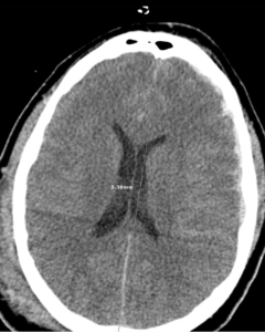
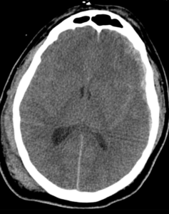
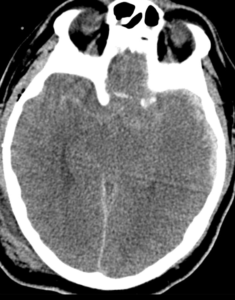
Imaging: CT of the head (see figures) showed subarachnoid hemorrhage with left frontal and temporal subdural hemorrhage, effacement of the suprasellar cistern, and effacement of the 3rd and 4th ventricles. In addition, CT studies showed a left frontal/temporal and parietal hematoma with mass effect and cerebral edema causing a 5.38-mm left to right midline shift

Room 9 Treatment: He required atropine push and nicardipine infusion (blood pressure was allowed to remain elevated to ensure continued brain perfusion for a target of SBP ~ 180. An arterial line, central venous catheter, and external ventricular drain were placed for fluid and medication administration and ICP management while the OR was prepped. Emergent treatment for herniation syndrome included endotracheal intubation with mild hyperventilation, 30 grams of IV Mannitol, HOB at 30 degrees, and EVD. Longer term treatment options include hypertonic solution of 23% (weight/volume) sodium chloride (NaCl) and after room 9 measures left-sided decompressive craniectomy.

Clinical course: Postoperatively, an external ventricular drain (EVD) was placed; the initial intracranial pressure (ICP) was 14 mmHg. The patient was examined postoperatively and his GCS was 5T, with bilaterally reactive pupils, and positive corneal reflex in the left eye. CT of his head showed improvement of midline shift and the ventriculostomy catheter tip was found to be in the proper location in the frontal horn of the right lateral ventricle  The patient was then started on 3% NaCl continuous infusion. ICP and cerebral perfusion pressure (CPP) displayed normal values at 3–4 and 70–75 mm Hg, respectively, for the first 24 h with the EVD open at 10 cm H2O.

His clinical course was complicated by VAP requiring antibiotics and bronchoscopy, later developing C Diff infection. He underwent tracheostomy and g-tube placement. His GCS examination improved to 10T, for which he was started on a neurostimulator drug amantadine. Ultimately, the patient was discharged to rehab.

A follow-up visit three months later revealed the patient was living at home with his mother. In the interim, his tracheostomy and gastrostomy tube had been removed. His major neurologic sequelae were transcortical motor aphasia and mood disorder. His GCS was 13 (E4, V3, M6). Ultimately his craniectomy defect was corrected surgically with a bone flap.

Teaching Points: When predicting mortality and unfavorable outcome following TBI, exam, laboratory, and imaging findings can be used together by utilizing the CRASH and IMPACT calculators. An unfavorable outcome is described as death, vegetative state, or severe disability. Here, we describe a patient who presented with a GCS of 3, bilaterally fixed pupils, and CT findings of subarachnoid bleeding, midline shift, subdural hematoma, effaced 3rd ventricle, effaced 4th ventricle, and effaced basal cisterns. Therefore, according to the CRASH calculator, which takes into account country, age, GCS, pupil reactivity, and CT findings, he had a 14-day mortality risk of 91.8% and a 95.7% chance of unfavorable outcome at 6 months

His pertinent laboratory studies, which are utilized along with exam and imaging findings in the IMPACT calculator, revealed an initial glucose concentration of 260 mg/dL and a hemoglobin concentration of 15.4 g/dL. Using the IMPACT calculator, at 6 months, the patient’s predicted probability of mortality was 62% and the probability of an unfavorable outcome was 77%.

He left our facility bedbound, ventilator- and tube feed-dependent, and in a minimally conscious state with a GCS 10T. Yet despite all this, he had a favorable recovery. Within 1 year of discharge, he was able to live at home, interact, and go shopping with his mother, walk, feed himself, and perform simple chores and ADLs

This is a poignant reminder that the variability between individual patients makes prognosticating after traumatic brain injury difficult and uncertain. This case shows that severe caution should be taken when using prior studies to make medical decisions about individual patients. Treatment of traumatic brain injuries is complex and should continue to evolve with evidence-based medicine. Improvement in outcome is not based on 1 intervention; rather, it is the additive effect of multiple interventions. In addition, the initial EMS response and ER Room 9 interventions along with later daily multidisciplinary rounds with Neurocritical Care, Trauma Critical Care, Infectious Disease, Pharmacy, Respiratory Therapy, Physical Therapy, Occupational Therapy, Social Services, Chaplain Services, and Dietary Services provided optimal medical management in a team-based approach. 

Wisdom from Dr. Mattu and Dr. Coleman

Here is some bonus material from Essentials of EM/ LITFL. A couple of lectures from Dr. Mattu from some of his favorite topics. It includes a lecture reviewing some of the hyperkalemia stuff we went over today.

Primum non killem


Also, I received a reply with some additional info from Dr. Coleman regarding epiglottitis and a sign called the vallecula sign which sometimes accompanies the thumbprint sign on a lateral neck x-ray.

From Dr. Coleman:

“Last week you had mentioned the radiographic soft issue sign on a lateral neck, the thumbprint sign as a good indicator of epiglottis. There are a few descriptions with 80-90 % sensitivities of a vellecula sign on the lateral soft tissue x-ray.
I have observed this sign, and can’t say that it was better or worse PPV or NPV that the epiglottis thumbprint, but in any instance that I am worried enough to obtain a soft tissue lateral of the neck, either awaiting for ct scan of unable to obtain e.g.patient can’t lay supine ( secretions, airway fear or comfort ) so a lateral neck is one of a limited choice, Having an additional sign has been helpful. Essentially the X-ray sign is a blunting of the sharp angle the epiglottis makes with the hypopharynx, where one is aiming for with a Macintosh blade. It would make sense that this space would reflect some early swelling, in an infectious process of the epiglottis.”
http://www.texasmedicalspa.com/assets/description-and-evaluation-of-the-vallecula-sign_a-new-radiologic-sign-in-the-diagnosis-of-adult-epiglottitis.pdf
Look up some images of the vallecula sign if you get a chance.
As always, thank you for all that you do!

Dislocation Video Links

All,

As promised during my lecture. See below for the links to the videos I referenced as well as a few others for joint reduction techniques. Hope these help. Also, I’m open to any feedback you have regarding the talk: things you liked, didn’t like, would like to see more of. Thanks.

https://www.youtube.com/watch?v=UxUhW4Zac74 Whistler Technique Hip Reduction Video

https://www.emrap.org/episode/reduction/reduction Multiple Techniques Hip Reduction

https://www.emrap.org/episode/hipreductionby/hipreductionby East Baltimore Lift- Hip

Larry Mellick Posterior Shoulder Reduction: https://www.youtube.com/watch?v=KRCqVekNEKc

Larry Mellick Inferior Shoulder Reduction https://www.youtube.com/watch?v=C8Irt39KBgk

Shorter version of Mellick’s Posterior Reduction w/o sound https://www.youtube.com/watch?v=MWb1OKkDDwE

Larry Mellick: 10 ways of reducing shoulder (10min) https://www.youtube.com/watch?v=HtOnreM7heg

Larry Mellick: Intra-articular injection/Ant Shoulder Reduction https://www.youtube.com/watch?v=HzROgg-HWPk

Larry Mellick: Davos Technique Shoulder Reduction https://www.youtube.com/watch?v=u2MsnjVNoPM

Jess Mason EMRAP: good explanation of Inf Shoulder Reduction https://www.youtube.com/watch?v=k_ORI51luFI

https://coreem.net/core/true-knee-patellar-dislocations/ Nice little summary on knee/patella dislocations

Larry Mellick Knee Dislocation: https://www.youtube.com/watch?v=aN7zDxtyHy8

Not Actual patients but good 2 minute video on techniques/exam: https://www.youtube.com/watch?v=vdrfY3K7yR4

https://www.youtube.com/watch?v=A91TWNbSEOQ – Silent Posterior Elbow Reduction Video

https://www.youtube.com/watch?v=IcrmMAxLnp8 – Interlocking Hands Technique for Posterior Reduction

https://www.youtube.com/watch?v=s9GVdF6v9xQ Posterior Elbow Reduction- 2 providers