![Room9er ["Room Niner"]:](https://room9er.com/wp-content/uploads/2020/03/cropped-Screen-Shot-2020-03-08-at-3.16.16-PM.png)
Alex Bequer, MD; Kahra Nix, MD. Peer Review Jeff Baker, MD
Emergency Medicine (EM) physicians are already familiar with the Focused Assessment with Sonography in Trauma (FAST) to rapidly identify intraperitoneal or pericardial free fluid and guide time-sensitive decisions. [1,2]There is an image processing error that EM physicians should be aware of as it can be confused with intraperitoneal free fluid.
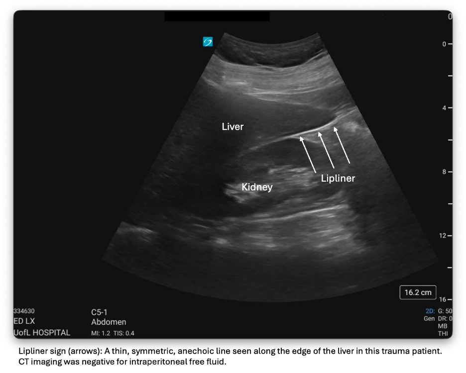
Modern ultrasound machines and software increasingly strive for sharp, high-contrast images, relying on post-processing in order to improve border definition and image clarity. These adaptions are enacted to prevent speckling and result in clearer, crisp images. While these features enhance visualization, they can also introduce other limitations in image interpretation. One example is the “lipliner” which appears as a thin, symmetric, anechoic line that can be seen along the edge of solid organs (see attached image). The lipliner can be found along the caudal edge of the liver and at the splenic tip, precisely where free fluid is expected on a positive FAST examination, thereby creating the potential for false-positive interpretations. [3] Unlike true free fluid, which typically forms a wedge-shaped, dependent collection that tracks into tissue planes, the lipliner outlines the solid organ margin itself. It is a result of real-time adaptive filtering, rather than anatomy or pathology, and because it is a mathematic result, the lipliner is not technically a sonographic artifact. [3]
It is important for EM physicians to recognize and understand the lipliner. Machine vendors and POCUS leaders are working on machine presets that may preserve image quality and minimize the appearance of the lipliner. Consider looking again and adjusting probe positioning to clarify if the anechoic area is wedge-shaped and extending into potential spaces. Consider serial FAST exams. [4]
In addition to other well-known mimics of free fluid, physicians should expect to encounter post-processing errors introduced by modern ultrasound advancements designed to improve image clarity and usability. Understanding how these technologies influence image appearance is critical to accurate interpretation and informed clinical decision-making. Watch out for the lipliner when interpreting FAST scans on your next shift.
References
1. Patel NY, Riherd JM. Focused assessment with sonography for trauma: methods, accuracy, and indications. Surgical Clinics of North America. 2011;91(1):195–207.
2. Rozycki GS, Ochsner MG, Feliciano DV, et al. Early detection of hemoperitoneum by ultrasound examination of the right upper quadrant: a multicenter study. Journal of Trauma. 1998;45(5):878–883.
3. Parker MA, Hicks BG, Kaili M, et al. The lipliner sign: potential cause of a false positive focused assessment with sonography in trauma (FAST) examination. Journal of Emergency Medicine. 2024;67(6):e553–e559.
4. Ferre, R. M., & Stolz, L. A. (2025, March 13). Lipliner artifact review. American College of Emergency Physicians, Emergency Ultrasound Section. https://www.acep.org/emultrasound/newsroom/march-2025/lipliner-artifact-review