Can you give Vasopressors through a PIV?

I looked back and couldn’t find a post about this topic in the last year or so but forgive me if it has already been posted. I have been following R.E.B.E.L. EM for a few months now  and I would recommend it to everyone who has the time and wants quick summaries on the latest EM literature.  They have short written summaries of papers including pros/cons of the study and what they feel are the most important take away points. It was founded by Salim Rezaie with Rob Rogers, Matt Astin and Anand Swaminatham serving as editors.

Occasionally they will have a “mythbuster” post looking at common myths in the ED and the most uptodate literature available on the topic. (their latest mythbust is on “safe” glucose levels before ED discharge but I digress)


Back in May they reviewed the safety of vasopressors through a PIV. The topic paper was titled “A systematic review of extravasation and local tissue injury from administration of vasopressors through peripheral intravenous catheters and central venous catheters

What the review found was very interesting: of 318 events, 204 results in local tissue damage, 114 were only extravasation events and 7 events involved the use of a CVC (so clearly not completely safe). Interesting, of the 204 local tissue events 85.3% involved PIV distal to the antecubital fossa and 96.8% involved administration of >4hrs.

REBEL EM’s take away points were:

In critically ill patients, with hemodynamic instability, vasopressor infusion through a proximal PIV (antecubital fossa or external jugular vein), for <4hours of duration is unlikely to result in tissue injury and will reduce the time it takes to achieve hemodynamic stability.

What I feel like this means for us is simple: If you have a crashing, hypotensive patient who needs a pressor without a CVC but good proximal PIVs, start the pressor immediately, stabilize the patient as best you can, then take the time to properly place a CVC.

Continue to monitor the PIV until it can be switch to the CVC and stop the pressor immediately if there is any suspicion for local extravasation. I am sure this will make some people nervous but I think this is better then placing a “crash line” that is less then sterile which will expose an already ill patient to infection or other complications secondary to a hastily placed CVC.

I highly recommend read their review and how they came to this conclusion along with their other posts. I have included the link to this study at the bottom.

http://rebelem.com/mythbuster-administration-of-vasopressors-through-peripheral-intravenous-access/

Name that Disease?

55 y/o  F with hx of HTN,COPD and recurrent indurative lesion on her left foot, last event was a yr or so ago, presented to the ED for worsening pain and increased size of her lesion for the last few wks now. No fever, chills, or fatigue. On exam, the cutaneous lesion is mildly tender and erythematous, non-fluctuant, no warmth noted. Pt is immunocompetent.

img_7436


/

/

/

/

/

/

/

/

/

/

/

/

Answer:

Pyoderma Gangrenosum.  Take home point is to not I&D this lesion. It is not an abscess. It is a rare autoimmune disease that affects pts in their 40s-50s. These pts will have hx of other autoimmune diseases–lupus, crohns etc.  An I&D would lead to phenomenon known as pathergy,  the formation of new lesions following a trauma.

Tx: High dose steroids and pain meds. Refer to podiatry. Pt in this case was already well known to podiatry on arrival and was discharged with steroids and pain meds after podiatry consult in the ED.

Paucis Verbis Cards “In Few Words”

Just in case people are not familiar with the free PV review cards as posted by ALiEM (more than 150), I have linked to a particularly good one dealing with algorithms for rash management: paucis-verbis.  These were also highlighted as very useful in the ACEP Now, Nov 2016; and back in 2013 they were brought up by Buckingham on Room9ER.  I have attached photos of the rash algorithms below, might be a potentially very applicable Norton Children’s reference.

screen-shot-2016-11-23-at-7-35-59-pm screen-shot-2016-11-23-at-7-36-16-pm screen-shot-2016-11-23-at-7-36-48-pm screen-shot-2016-11-23-at-7-37-02-pm


Approach to the Fussy Infant

There are many different types of challenging patients that we all dread seeing when they pop up on the board. Whether the patient’s chief complaint is headache, back pain, or pregnant female with abdominal pain. Another very challenging patient presentation is the crying infant. The differential when evaluating a crying infant is broad. In this post I will include a list of differential diagnosis to consider based on organ system and then a patient I had that presented in this way.

CNS- Meningitis, epidural hematoma, subdural hematoma, hydrocephalus

HEENT- Skull fracture (accidental or non- accidental trauma), ocular foreign body, corneal abrasion, otitis media, nasal foreign body.

Cardiovascular- SVT, myocarditis, congestive heart failure

Pulmonary- foreign body in airway, bronchiolitis, pneumonia

GI- Malrotation/volvulus, pyloric stenosis, appendicitis, gastro-esophageal reflux, intussusceptions, anal fissure

GU- Testicular torsion, UTI, incarcerated inguinal hernia, soap vaginitis, phimosis, paraphimosis

Musculoskeletal- Fracture, septic arthritis, dislocation, hair tourniquet

My patient presented as a 1 month old male with his Spanish speaking Hispanic parents. Mom stated that he has been crying consistently for the past 4 days. She does not remember a specific time when the crying started but she states it has not improved.

Mom is breastfeeding the baby and denies any dietary changes. The baby was full term and mom had no complications with the pregnancy. Patient up to date on all vaccinations. Baby has been afebrile and per mom has not been lethargic. Baby is still feeding well and gaining weight appropriately and mom denies any projectile vomiting after feeds, denies any change in stooling, and notes good urine output. Baby lives at home with mom and dad and I have no red flags to suspect non-accidental trauma.

On exam he is overall well-appearing and does not appear to be in any type of distress: crying but consolable. He appeared to be healthy. He was interactive, tracking me with his eye movements, and did not appear to be meningitic or lethargic. Heart and lungs were unremarkable and abdomen was soft, nontender and non distended. GU exam (make sure you do this) was unremarkable. Baby was moving all extremities while lying on the oversized adult bed in the middle of the hallway and on inital exam did not appear to have any outward bruising or signs of trauma.

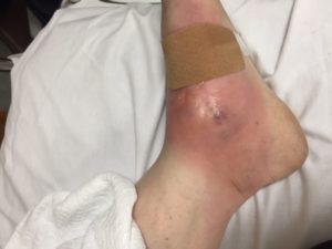
Upon removing the patient’s socks I noticed something odd on his second toe of his right foot. Just distal to his PIP joint he had a circumferential red line. As soon as I started to examine that toe his crying increased substantially. On further examination he had a hair tourniquet that had eroded its way all the way down to the bone of the middle phalanx of the second toe.

At this point the baby was still in the hallway and we took him to Room 9 to attempt to try any remove it. I am sure you can imagine how awesome this was on a crying kicking 1 month old. We attempted to unwind the hair but ultimately were unable to do so as it was just to deep into the tissue. I called Kosair and the patient was transferred and I do not yet follow up on the final outcome.

This just re-enforces the importance of a good head to ahem, toe physical exam on patient’s that are not straight forward. Mom had been with the infant 24/7 for the past 4 days and had not noticed this; not to mention who knows when the hair tourniquet actually started. Just something to keep in mind and hopefully this helps next time you all have to examine a crying baby.


Central Line Insertion Choice

All,
I know this came up during conference today so thought I’d send the article I think was cited. At least this is the one I found from EMRAP, below is their summary of this article. Long and short of it, complication rates are really low, when done in sterile fashion in a controlled environment.
This was done in the ICU, not in the emergency department.
Overall they didn’t say that one site was absolutely the best.

nejm-2015-central-line-site-complications

Take Home Points
No central line site is superior.
Femoral lines are fastest and most successful. Subclavian lines have a lower infection risk but higher rate of pneumothorax.

Parienti, JJ et al. Intravascular complications of central venous catheterization by insertion site. N Engl J Med. 2015 Sep 24;373(13):1220-9. PMID: 26398070

Bottom line: no line was superior. Femoral lines were the fastest to place and had the highest success rates. Subclavian lines had the lowest infection risk but had a higher rate of pneumothorax.

A patient needs central line access. Which should we choose? Which is best? There are multiple complications; infection, mechanical complications like artery puncture or pneumothorax and thrombotic complications.

The authors of this study conducted a randomized, controlled trial in 10 French ICUs. They enrolled adult patients with at least two accessible sites. Patients with all three sites accessible were randomized in a 1:1:1 fashion while those with only two sites were randomized in a 1:1 fashion. The doctors had all performed at least fifty central lines. However, they were all aware of the study and probably tried harder to reduce complications.

They looked for symptomatic clots and/or infection from the time of insertion up to 48 hours after removal. This was a large study; 3471 catheters were placed in 3027 patients. Catheters were assigned to a randomly assigned site and side; placement was successful approximately 91% of the time. 85% of subclavian lines were successfully placed, 91% of the jugular lines were placed and 95% of the femoral lines were placed. Femoral lines were most successful and subclavian lines were least likely successful.

Placement of femoral lines was also more rapid, by about a minute.

The primary outcome was a composite of infection, symptomatic clot and mechanical complications such as pneumothorax and bladder puncture. The jugular line performed the worst followed by the femoral line, then subclavian line. However, it is important to look at the individual components.
For mechanical complications, the subclavian line performed the worse. 2% had a complication versus 1.5% of jugular lines and less than 1% of femoral lines.
All lines were fairly low for symptomatic clots; 0.5% for subclavian, 1% for jugular and 1.4% for the femoral group.
In terms of infection, the subclavian group was the lowest (0.5%). The highest? Surprisingly, jugular lines had a 1.4% rate of infection versus 1.2% in the femoral line group.
Overall, there were fairly low rates of complications. These were performed in very sterile conditions in the ICU. These were not placements in crashing or coding ED patients.

Placement of central lines, including femoral lines, when done carefully under sterile conditions has a low rate of complications. This article does not identify one superior line placement.

Studying Core Texts

I’ve recently had several of the interns asking me about studying. I think all of us should be studying one of the core texts: Rosen’s, Tintinalli’s, or Harwood-Nuss. My own preference is for Tintinalli’s for a number of reasons. It is the most direct, in terms of what do I need to know to take care of a patient with this condition. It is also slightly more comprehensive than Rosen’s. I also love that it has three versions: Emergency Medicine: A Comprehensive Study Guide; Emergency Medicine Manual (with a new version coming out soon); and Emergency Medicine: Just the Facts. For those that don’t know, the manual is a shortened version of the big book, still in paragraph form.  Just the Facts is an outline version. I use the big book as my primary resource, the manual when I just can’t make myself read the renal chapter, and Just the Facts as a quick review.

Rosen’s is a wonderful book, extremely well written, and something I plan to read after residency. Hawood-Nuss is too simplistic, leaves out too much of what a good ER doctor needs to know, and in my opinion is only suitable for NPs and PAs.

If you are trying to decide which book works for you, pick a couple of chapters of each book, read the chapters and see what works better. Notice that I say a couple chapters, because each chapter is written by different authors. You don’t won’t to stick with less than ideal book, just because one chapter was written beautifully.

A resource I recently discovered and have been enjoying is CrackCast. It is written by a Canadian group and they publish a new lecture each week going over a single chapter of Rosen’s, in order. Now even though I am a Tintinalli’s man and the material is slightly different, core content is core content. The lectures are wonderful.

CRACKCast

Something else wonderful about this group is their belief in spaced repetition. The group publishes flashcards for every lecture that they do.

Flashcards

For our interns, here are a couple previous blog posts about learning and spaced repetition:

Science of Learning

Spaced Repetition


Managing Migraine

As mentioned on R and R in the Fast Lane. This article by Friedman is a welcomed update to evidence-based migraine management. Some people love treating migraine patients, some hate it. But we all have our cocktails we believe in.

I am a fioricet/neurontin/IM compazine, escalating to IV compazine/benadryl/decadron/toradol OR if they want to drive home IV MAG/decadron/toradol … kind of guy.

This article starts with criteria for delineating migraine from other headache forms. Then provides a succinct algorithm for treatment, starting with reglan or compazine +/- benadryl, then another dose plus toradol, then dihydroergotamine, then occipital nerve blocks (very fun), then as a last resort, opioids. I would encourage you to attempt a few other methods before the blocks and especially before the opioids.

Many other medications can be used (keppra, depakote, propofol, etc). But this is a solid overview of the EM approach. Also of note, see the Oct 2016 EM-RAP paper chase of the reglan +/- IV fluids in migraine article which showed no real benefit to the addition of IVF.

Stop Traffic

Hey guys some of you were lucky enough to hear my friend Olivia Mittel and her guests educate our group in didactics. She presented information on human trafficking. This is a real issue and many of us have likely discharged a trafficking victim back into a terrible situation. Please read this Annals of EM article for more information very relevant to EM doctors.

Headache in a post-neurosurgical patient

Earlier this month while moonlighting I had an approximately 40yM present for a headache (9/10 pain) for 3 days.

I know, this is an everyday occurrence.

But in his case he had a craniotomy with removal of a meningioma 1 month prior. He also noted 2 weeks of swelling on the left side of his head along the surgical scar.
ROS: denied fevers, chills, changes in vision, weakness, numbness, or tingling, etc.

PE: VSS, HEENT: Left side of head extending from his surgical scar and wrapping around to even under his L eyebrow was swollen and firm. NEURO: WNL

So, I know something isn’t right and my guess is that he either had a bleed or infection associated with his surgery. I order a CT head. For his headache I give him a migraine cocktail (IVF, compazine, and benadryl, minus the toradol).

img_1534

img_1535

After I see the CT images, I rush to check on the patient as the CT obviously shows quite a bit of midline shift and the patient states his headache is drastically improved (2/10 down from 9/10). Apparently migraine cocktails work on all causes of headache.

I consulted Neurosurgery who promptly admitted the patient to the ICU with plans to go to the OR. Approximately 45 minutes later, after the NES nurse practioner has seen the patient and he is getting packed up to go to the ICU, the radiologist calls to notify me of the CT findings.

This is a reminder to ALWAYS look at CT images yourself, especially if you’re expecting a life threatening finding.

Lactate

Bosse’s favorite molecule (and lab test), lactate has a sordid and confusing history. This article in Emergency Medicine News sheds some light on lactate and its role in disease (or health). The author (Dr Paul Marik) is board certified in internal medicine, critical care, neuro-critical care and nutrition.

It may take a couple reads but this article has a succinct, common sense approach to understanding lactate production and use by the body. You may not be able to apply this knowledge on your next patient encounter, but physiology is fun right?

Chest pain?

Yesterday, 10 minutes before the end of a very busy shift in the middle of nowhere:
Nurse hand’s me an EKG for the mom of one of our best young medics. Ischemic STD in II, III, aVF, V5, V6. No STE. Maybe some LVH. No other EKG on file, she hasn’t been here before. I get up to see her right away.
In the room is a 50 yo lady in severe distress. BP 250/140. Describes “tearing” L sided CP radiating to her upper back. I ask her if she has a family hx of sudden death or aneurysms, her son says yes. No SOA. No N/V. Lungs are clear. Can’t palpate pulses in her feet. Slightly obese. She is sweating. There isn’t a CT surgeon in this county; I am sweating, too.
The nurse gets CT ready and I start the ball rolling for big IVs, blood, helicopter, etc. She gets dilaudid 1 mg then 0.5 then 0.5 again for pain and hopefully BP control. I tell everyone she’s now the ED’s top priority. The family of another patient grabs me twice in the hallway and asks why their mom, who fell, hasn’t gotten her home dose of lisinopril yet.
~30 minutes from door to imaging – CTAs = no dissection. Great, I have some time. Pain is well controlled now and BP 150s/80s. Repeat EKG w ischemia resolved completely, normal. Hypertensive emergency? She has a bit of a headache so I scan her head because we’re in CT, but it’s not useful due to the residual contrast. Trops, CBC, CMP, urine, tox, etc. all negative. Now looks great and feeling much better. Wants to go home.
Her son, her nurse, and I spend a long time convincing her she needs to stay overnight. She doesn’t want to be admitted, as she’s starting a new job Tuesday and can’t miss it. Eventually, she agrees to stay. Then we have to convince her to be transferred, since ED MD is the only MD in house overnight and she had me straight terrified. Reluctantly, she agrees to the transfer as long as her son will bring her home Monday night, no matter what. Hospitalist at the local mothership accepts readily. At that hospital, she’d had a negative stress 8 months ago and NES had placed her shunt (pseudotumor, she has a lot of headaches); hospitalist will consult cards and NES when she gets in.
I arrive back to work this morning to find out she’d made it upstate only to be transferred again overnight. She’d been flown to a bigger center after repeat CT head demonstrated SAH. She had 2 aneurysms, 1 was bleeding, both were definitively addressed. 24 hours after walking in the door, at the time of this writing, she is extubated and thriving.
Found this article from 1988 with a similar presentation, but I can’t say I’d ever heard of it before. Something else for your differential. Really consider that admission for observation when something seems wrong, even if you don’t know what it is.
Going to change my drawers now.